
Image
|
Figure Caption
Fig. 6
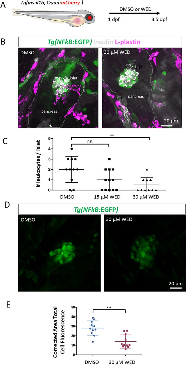
Wedelolactone treatment ameliorates immune-cell infiltration and reduces islet NF-κB signalling activation.(A) Schematic of the experimental approach. Tg(ins:il1b);Tg(NF-kB:GFP) embryos were treated with DMSO or wedelolactone (WED) from 1 to 3.5 dpf. At 3.5 dpf, the presence of L-plastin-positive leukocytes in the islet region was quantified. (B) Representative confocal images of Tg(ins:il1b);Tg(NF-kB:GFP) embryos treated with DMSO or 30 μM wedelolactone. Whereas the leukocytes have infiltrated the islet in DMSO controls, there is a reduction in islet-associated immune cells following wedelolactone treatment. (C) Quantification of the number of L-plastin-expressing cells in the islets of DMSO- and wedelolactone-treated larvae. One-way ANOVA with Sidak's multiple-comparison test; **P≤0.01; ns, not significant. Mean±s.d. (D) Representative confocal images of the islets of Tg(ins:il1b);Tg(NF-kB:GFP) embryos treated with DMSO or wedelolactone from 1 to 4 dpf. Wedelolactone-treated larvae show a visible reduction in NF-kB:GFP fluorescence intensity in the islet region at 4 dpf. (E) Quantification of corrected area total cell NF-kB:GFP fluorescence in the islet region following DMSO or wedelolactone treatment, showing a reduction in NF-kB:GFP fluorescence. Unpaired two-tailed t-test with Welch's correction, ***P≤0.05. Mean±s.d.
Figure Data
Acknowledgments
This image is the copyrighted work of the attributed author or publisher, and
ZFIN has permission only to display this image to its users.
Additional permissions should be obtained from the applicable author or publisher of the image.
Full text @ Dis. Model. Mech.