IMAGE

Fig. 6

ID
ZDB-IMAGE-200227-7
Antibodies
Source
Figures for Chen et al., 2020
Image
Figure Caption

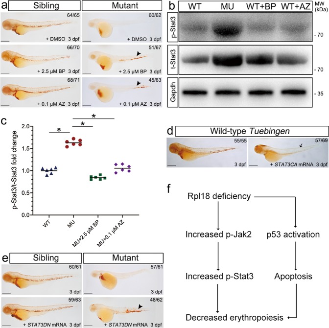
Fig. 6 Inhibition of the phosphorylation of Jak2 or Stat3 rescued hemoglobin level in <italic>rpl18</italic> mutants.

arpl18 sibling and mutant embryos were treated with BP (T3708, STAT3 inhibitor) or AZ (T6309, JAK2 inhibitor) (N = 5). At 3 dpf, o-dianisidine staining of mutant embryos showed obvious recovery with BP (2.5 μM) treatment, and mutants treated with AZ (0.1 μM) also be seen partially restored (arrowhead). b The total amounts of Stat3 (t-Stat3) and the phosphorylation status of endogenous Stat3 protein (p-Stat3) was evaluated in rpl18 siblings and mutants. The protein levels of t-Stat3 and p-Stat3 had a significant increase in mutants, compared with wild-type (WT) embryos at 30 hpf. Both the t-Stat3 and p-Stat3 were dramatically decreased in Rpl18-deficient embryos treated with BP or AZ separately, as shown by western blotting. c The scatter diagram represented quantification of western blots. The results of three groups of experiments (twice technical repeated for each group) were statistically analyzed, *P < 0.05. d The o-dianisidine staining displayed that most of erythroid cells disappeared (arrow) in wild-type embryos after injection the STAT3CA (constitutively active STAT3 with the double mutation A661C N663C) mRNA, compared with sibling embryos at 3 dpf (N = 3). e Erythrocytes were partially rescued in rpl18 mutants that had been injected with STAT3DN (dominant-negative STAT3) mRNA at 3 dpf (N = 3). Black arrowhead points to the recovery of erythrocytes. f Model of the defective erythroid maturation in Rpl18 deficiency through increased phosphorylated Stat3 expression. All scale bars represent 250 μm.

Figure Data
Acknowledgments
This image is the copyrighted work of the attributed author or publisher, and ZFIN has permission only to display this image to its users. Additional permissions should be obtained from the applicable author or publisher of the image. Full text @ Cell Death Dis.