IMAGE

Figure 5

ID
ZDB-IMAGE-200226-35
Source
Figures for Chávez et al., 2020
Image
Figure Caption

Figure 5

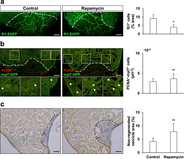
Rapamycin treatment affects zebrafish cardiac regeneration. Rapamycin-treatment affected angiogenesis upon amputation, causing a negative effect in the re-vascularization rate of the regenerating zebrafish ventricle at 7 dpa (a, fli1a+-covered area expressed in relation to the regenerating ventricle area); yet, the same treatment had a specific positive effect over cardiomyocyte proliferation at 14 dpa (normalized by the area considered for the quantification) (b). Still, sustained rapamycin administration impaired zebrafish cardiac regeneration, as shown by the AFOG-staining of amputated treated and control hearts at 28 dpa, which allows to visualize the remains of fibrin (red)- and collagen (blue)- provisional extracellular matrices, as well as the uninjured/restored cardiac muscle (yellow/brown) (c). The quantification of the non-regenerated area (normalized to the total ventricle area) showed that treatment with rapamycin for up to 14 dpa caused a significant impairment of the regenerative outcome of the zebrafish heart. Scale bar represents 100 µm in (a,b) and 50 µm in (c). fli1a+: Control N ≥ 8, Rapamycin N ≥ 7; PCNA+-myl7+: Control N ≥ 8, Rapamycin N ≥ 9, AFOG-staining: Control N ≥ 10, Rapamycin N ≥ 12. *p ≤ 0.05, **p ≤ 0.001.

Acknowledgments
This image is the copyrighted work of the attributed author or publisher, and ZFIN has permission only to display this image to its users. Additional permissions should be obtained from the applicable author or publisher of the image. Full text @ Sci. Rep.