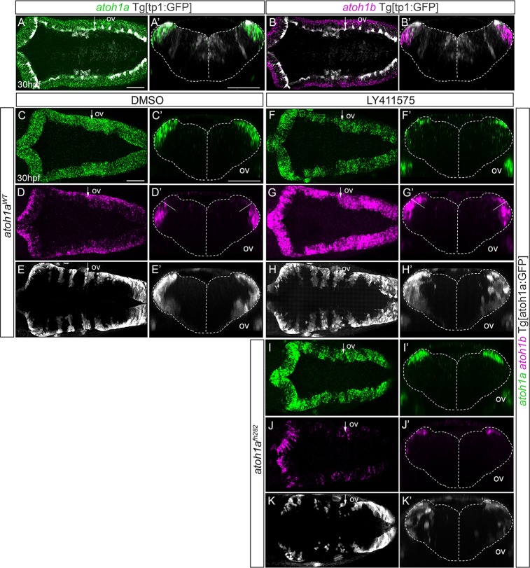
IMAGE

Fig 7

ID
ZDB-IMAGE-200225-7
Genes
Source
Figures for Belzunce et al., 2020
Image
Figure Caption

Fig 7 Notch-signaling regulates the transition of <italic>atoh1a</italic> cycling progenitors towards <italic>atoh1b</italic> committed cells.

A-B) Whole mount double in situ hybridization with atoh1a (green) and atoh1b (magenta) in Tg[tp1:GFP] embryos (readout of Notch-activity in white). A’-B’) Reconstructed transverse views of embryos displayed as dorsal views in (A-B) through the point indicated by the white arrow. Note that Notch-activity is restricted to the most dorsomedial tip of the hindbrain, corresponding with atoh1a cells. C-K) atoh1aWTTg[atoh1:GFP] (C-H) and atoh1afh282Tg[atoh1:GFP] (I-K) siblings were double in situ hybridized with atoh1a (green) and atoh1b (magenta) after treatment with DMSO (C-E, n = 10) or the gamma-secretase inhibitor LY411575 (F-H, n = 15; I-K n = 3). The atoh1a derivatives were followed by anti-GFP staining in white. C’-K’) Reconstructed transverse views of embryos displayed as dorsal views in (C-K) at the level indicated by the white arrow. Note how the atoh1b-domain expands at expense of atoh1a progenitors after blocking Notch-activity in wild type embryos, but not in atoh1afh282 mutants. A-D, F-G, I-J) Dorsal views of confocal MIP from dorsal stacks with anterior to the left. E, H, K) Dorsal views of confocal MIP from ventral hindbrain with anterior to the left. ov, otic vesicle. Scale bars correspond to 50 μm.

Figure Data
Acknowledgments
This image is the copyrighted work of the attributed author or publisher, and ZFIN has permission only to display this image to its users. Additional permissions should be obtained from the applicable author or publisher of the image. Full text @ PLoS One