IMAGE

Fig 3

ID
ZDB-IMAGE-200225-3
Genes
Source
Figures for Belzunce et al., 2020
Image
Figure Caption

Fig 3 Cell lineages and behavior of <italic>atoh1a</italic>-derivatives.

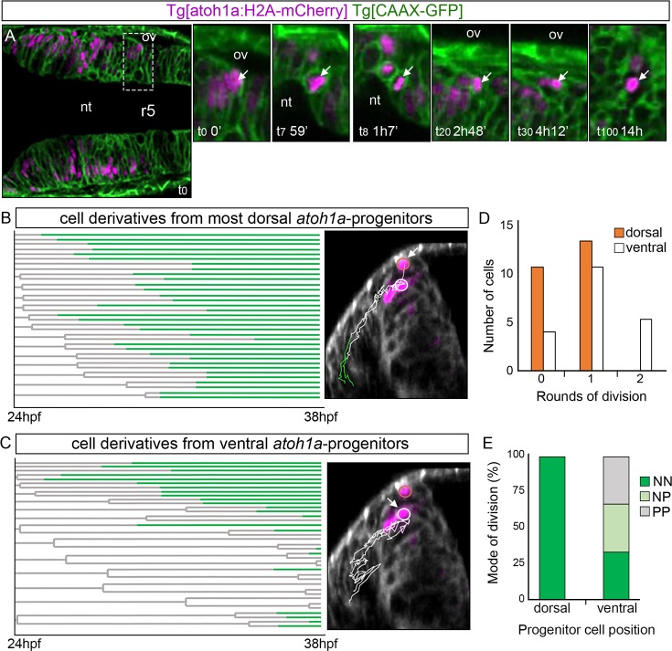
Tg[atoh1a:H2A-mCherry] Tg[CAAX:GFP] embryos were imaged from 24hpf during 14h, and information about cell position was acquired every 7min. A) Dorsal view of an embryonic hindbrain displaying atoh1a cells in magenta with anterior to the left. The inserts display magnified stills from the framed area in (A) at different times (see white arrow as example of a cell that was tracked from t0 to t100). Note the cell nucleus displacement towards the apical side before division (t8). B-C) Cell lineages from r4 and r5 atoh1a-progenitors located at different dorsoventral levels within the atoh1a domain; n = 22 in (B) and n = 17 in (C). Each line corresponds to a single cell that branches upon division. Lines are colored according to cell differentiation status: progenitors in grey and differentiated cells in green. The X-axis corresponds to developmental time. The right-hand images display examples of the trajectories of the atoh1a tracked cells (white arrow) on the top of the transverse views at t0 (24hpf). Cell trajectories are color-coded according to cell differentiation status: progenitors are in white and differentiated cells in green. Cells are considered differentiated neurons when they are within the neuronal differentiation domain. Dorsal most atoh1a cells are encircled in orange and ventral atoh1a cells are encircled in white. D) Histogram displaying the number of most dorsal (orange) or ventral (white) atoh1a:GFP cells that undergo different number of divisions over time. Note that atoh1a-cells that are more dorsally located undergo less division rounds (orange bars) than the ones in a more ventral position (white bars). E) Mode of cell division according to the DV position of the atoh1a-progenitor cells. NN, progenitors giving rise to two neurons; NP, progenitors generating one neuron and one progenitor; PP, progenitor cells that give rise to two progenitors. Note that most dorsal atoh1a cells give rise to differentiated cells in all analyzed cases (n = 22 atoh1a progenitors), whereas atoh1a cells more ventrally located employ the three modes of division (n = 17 atoh1a progenitors). nt, lumen of the neural tube; ov, otic vesicle; r, rhombomere.

Figure Data
Acknowledgments
This image is the copyrighted work of the attributed author or publisher, and ZFIN has permission only to display this image to its users. Additional permissions should be obtained from the applicable author or publisher of the image. Full text @ PLoS One