IMAGE

Figure 2.

ID
ZDB-IMAGE-200210-19
Source
Figures for Chhabria et al., 2018
Image
Figure Caption

Figure 2.

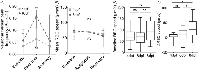
Neurovascular coupling in zebrafish develops after 6 dpf. (a) Quantification of frequency of peaks in (ΔF/Fo) as a measure of neuronal activation during the baseline, response and recovery time periods for 4dpf and 6dpf (n = 17 larvae/group). (b) Mean RBC speed during baseline, response and recovery time periods in the same animals as (a) (n = 17 larvae/group). (c) Mean baseline RBC speed in tectal vessels in 4, 6 and 8 dpf zebrafish. (d) Change in RBC speed (ΔRBC) in response to stimulus in 4, 6, and 8 dpf (n = 17 larvae/group). *p < 0.05, **p < 0.01. Data in (a) and (b) are mean ± s.d.

Acknowledgments
This image is the copyrighted work of the attributed author or publisher, and ZFIN has permission only to display this image to its users. Additional permissions should be obtained from the applicable author or publisher of the image. Full text @ J. Cereb. Blood Flow Metab.