IMAGE

Fig. 6

ID
ZDB-IMAGE-200205-23
Source
Figures for Simões et al., 2020
Image
Figure Caption

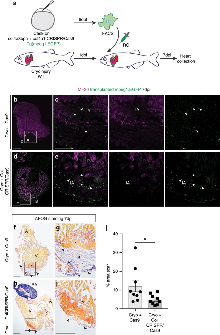
Fig. 6 Targeting <italic>col4a1</italic> and <italic>col4a3bpa</italic> in macrophages reduced scarring in zebrafish cryoinjured hearts.

a Schematics of adoptive transfer of GFP+ macrophages sorted from embryos injected with Cas9 only or col4a3bpa plus col4a1 sgRNAs/Cas9 and transplanted into a cryoinjured heart via retro-orbital (RO) injection. be Confocal imaging of anti-MF20 (fuschia) and anti-GFP (green) antibody-stained WT recipient hearts of Cas9 only (b, white boxes enlarged in c for detail) and col4a3bpa plus col4a1 sgRNAs/Cas9 (d, white boxes enlarged in e for high-magnification) transplanted macrophages (GFP+) collected 7 days post injury (dpi). Arrowheads point to GFP+ transplanted macrophages that are located in the site of injury (IA). Scale bar: 200 μm (insets showing high-magnification images, scale bar: 100 μm). Dotted line demarcates the MF20-negative injured area. Representative images of n = 3 per group. (fi) AFOG staining of representative images showing healthy myocardium (yellow), injured myocardium (orange) and collagen (blue). Excess scar tissue (arrowheads) at the periphery of the cryoinjured area (black asterisk) is seen in hearts transplanted with 6 days post fertilization (dpf) GFP+ macrophages injected with Cas9 only (f, with high-magnification inset in g). Hearts transplanted with col4a1+col4a3bpa CRISPR/Cas9 macrophages show reduced collagen staining (h, with high-magnification inset in i); red asterisk, myocardial dead tissue removed from the injury region. Representative images of n = 3 per group. j Quantification as a percentage of total section area (6 sections per heart) show significantly less scar fibrosis in the group of cryoinjured hearts transplanted with col4a1+col4a3bpa CRISPR/Cas9 macrophages (Cryo+Cas9: 12.18 ± 3.12%, n = 9 vs Cryo+Col CRISPR/Cas9: 4.91 ± 0.88%, n = 9; *p= 0.039; data are mean ± SEM, 2-tailed, unpaired Student’s t-test). Scale bar: 200 μm; high-magnification insets scale bar: 50 μm; A, atrium; V, ventricle; BA, Bulbus Arteriosus. Source data are provided as a Source Data file.

Acknowledgments
This image is the copyrighted work of the attributed author or publisher, and ZFIN has permission only to display this image to its users. Additional permissions should be obtained from the applicable author or publisher of the image. Full text @ Nat. Commun.