
|
Fig. 8
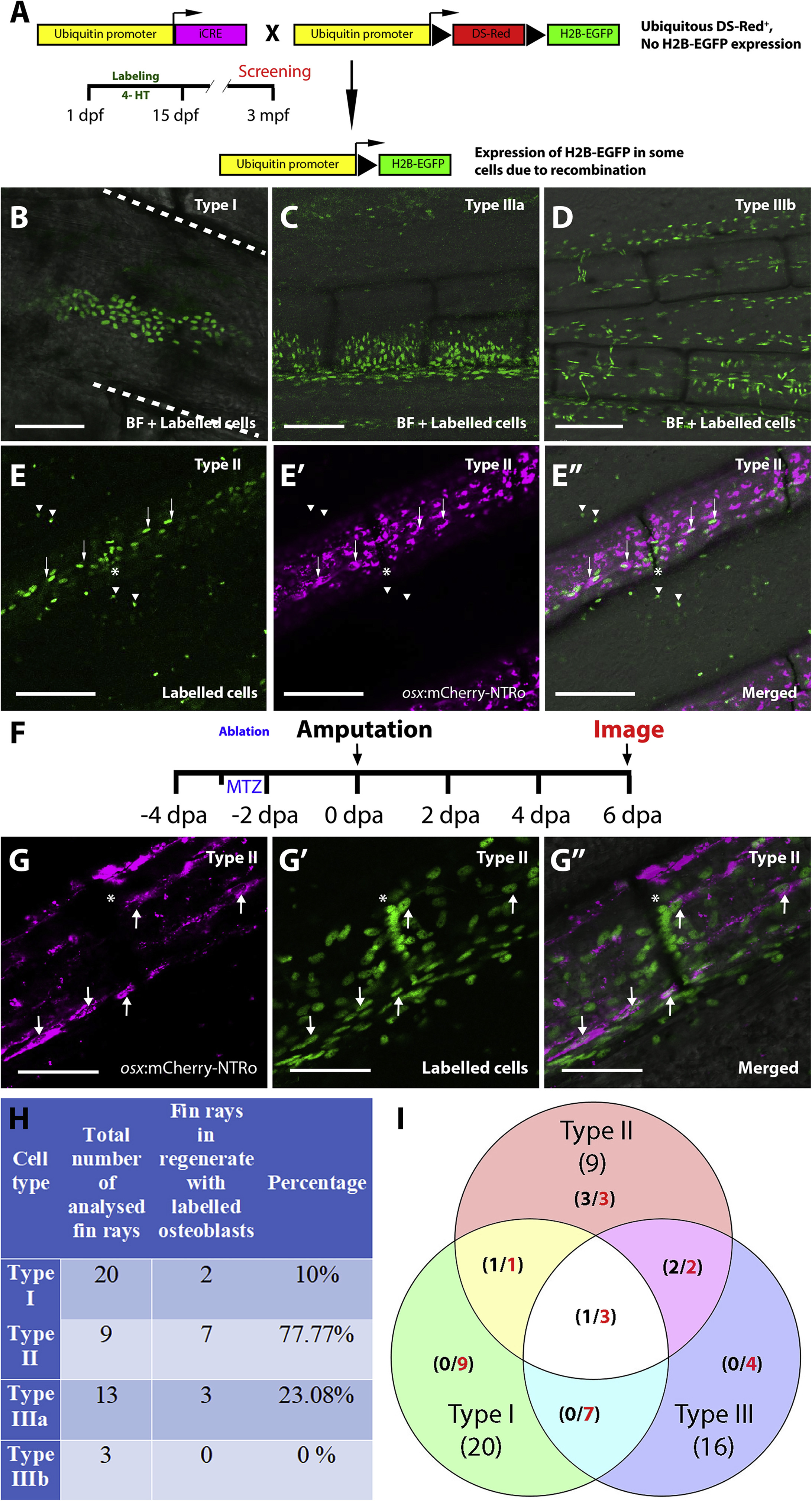
Lineage tracing of randomly labelled cells identifies sources for osteoblast regeneration. A: Strategy for labelling different cell lineages by 4-HT incubation at embryonic and post-hatching stages (1 – 15 dpf) and detection of labelled cells in the adult fin (3 mpf). Triangle represents loxP sites. B-E’‘: Confocal images of adult fins at 3 mpf showing labelling of different cell types (identities were predicted based on position and morphology; see Tu and Johnson, 2011): Type I (epidermal and blood cells, B), Type IIIa (intraray fibroblasts; C) and Type IIIb (endothelial cells; D). The panel in E shows Type II cells (osteoblasts) that co-express osx:mCherry-NTRo (E′), as evident in merged image (E”; EGFP, mCherry and brightfield merged images). Dashed white lines in B indicate borders of fin rays. Arrowheads indicate GFP signal outside the fin rays of unclear origin. Scale bars = 100 μm. F: Timeline for osx cell ablation, amputation and imaging of labelled cells in the regnerating fin. G: Representative confocal images showing osx:mCherry-NTRo expressing osteoblasts (G), lineage traced type II cells (G′), and merged image at 6 dpa. Note osx expression in some of the labelled cells (arrows). Asterisks label the intersegmental regions. Scale bars = 100 μm. H,I: Table (H) and Venn diagram (I) showing contribution of different classes of labelled cells (Types I to III) to the osteoblast lineage. Numbers in red indicate total number of fin rays positive for particular cell type, numbers in black indicate fin rays with contribution to osteoblasts (after ablation) per number of analysed fin rays.
Reprinted from Developmental Biology, 455(1), Dasyani, M., Tan, W.H., Sundaram, S., Imangali, N., Centanin, L., Wittbrodt, J., Winkler, C., Lineage tracing of col10a1 cells identifies distinct progenitor populations for osteoblasts and joint cells in the regenerating fin of medaka (Oryzias latipes), 85-99, Copyright (2019) with permission from Elsevier. Full text @ Dev. Biol.