IMAGE

Figure 1

ID
ZDB-IMAGE-200129-34
Genes
Antibodies
Source
Figures for Fallata et al., 2019
Image
Figure Caption

Figure 1

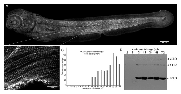
Mmp2 is expressed ubiquitously from early development and accumulates in a striated pattern within the skeletal muscle. (A) Composite confocal projections of a 72 hpf embryo stained with anti-Mmp2 exhibiting labeling throughout the embryo with notable accumulation in the skeletal muscle (scale bar = 200 µm). (B) High magnification view of a single confocal section through the trunk musculature (indicated by the inset) showing strong labeling of the myotome boundary (upper left corner), and striated staining in myofibrils (scale bar = 10 µm). (C) RNASeq data showing absolute abundance of mmp2 transcripts in embryos from fertilization to five days post-fertilization (dpf). (D) Immunoblot of whole embryo homogenates (350 µg per lane) made from 2 hpf (cleavage), 5 hpf (50% epiboly), 12 hpf (early somitogenesis), 18 hpf (late somitogenesis), 24 hpf (prim-5), 48 hpf (long pec), and 72 hpf (protruding mouth) embryos probed with anti-Mmp2. Immunoreactivity is detected in embryos after 12 h of development, with bands at the expected mobility for full-length Mmp2 (72 kD) and stronger bands at 44 and 20 kD, which combine to give the expected size for activated Mmp2 (64 kD).

Figure Data
Acknowledgments
This image is the copyrighted work of the attributed author or publisher, and ZFIN has permission only to display this image to its users. Additional permissions should be obtained from the applicable author or publisher of the image. Full text @ Biomedicines