
Image
|
Figure Caption
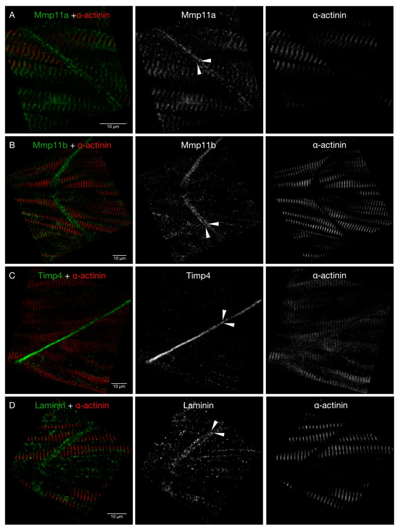
Figure 5
Timp4 and both Mmp11 paralogues are present in the MTJ at 28 hpf. High-resolution confocal micrographs of MTJs in the trunk skeletal musculature dorsal to the yolk extension of 28 hpf embryos stained with antibodies against (
Figure Data
Acknowledgments
This image is the copyrighted work of the attributed author or publisher, and
ZFIN has permission only to display this image to its users.
Additional permissions should be obtained from the applicable author or publisher of the image.
Full text @ J Dev Biol