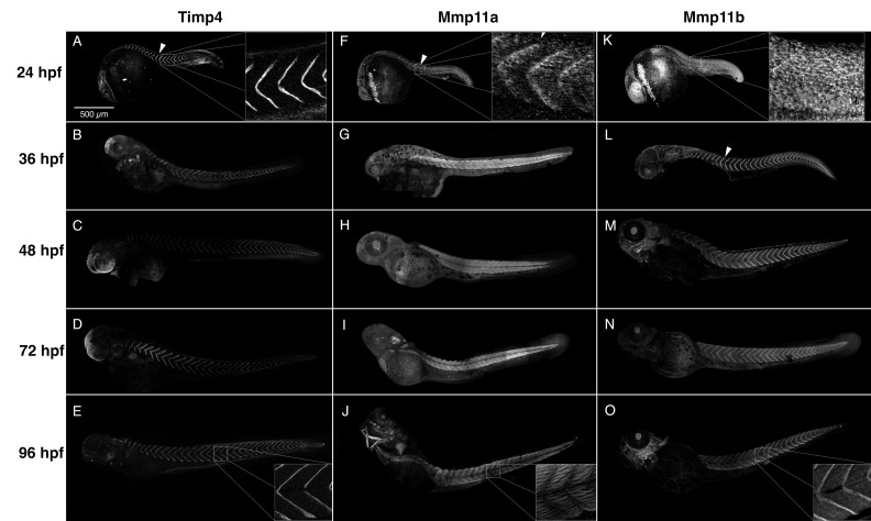
IMAGE

Figure 4

ID
ZDB-IMAGE-200125-59
Genes
Antibodies
Source
Figures for Matchett et al., 2019
Image
Figure Caption

Figure 4

Timp4 and Mmp11 paralogues accumulate dynamically during development and co-localize at the myotendinous junctions (MTJs). Composite confocal projections of 24, 36, 48, 72 and 96 hpf embryos labeled with antibodies against Timp4, Mmp11a, or Mmp11b. (AE) Timp4 immunoreactivity is abundant at the MTJ at all stages examined (arrowhead), and accumulates dynamically in ectodermal epithelia of the head, fin folds, otic vesicle, the posterior notochord and muscle attachments in the jaws and pectoral fins in later stages. (FJ) Mmp11a is present at the MTJs in 24 hpf embryos (F) (arrowhead) but becomes localized within muscle cells in later stages (G,H). (KO) Mmp11b becomes concentrated in MTJs at later stages (LO) (arrowhead). Insets show higher magnification views of MTJs dorsal to the yolk extension in 24 and 96 hpf embryos for comparison. Scale bar = 500 µm.

Figure Data
Acknowledgments
This image is the copyrighted work of the attributed author or publisher, and ZFIN has permission only to display this image to its users. Additional permissions should be obtained from the applicable author or publisher of the image. Full text @ J Dev Biol