
|
Fig. 6
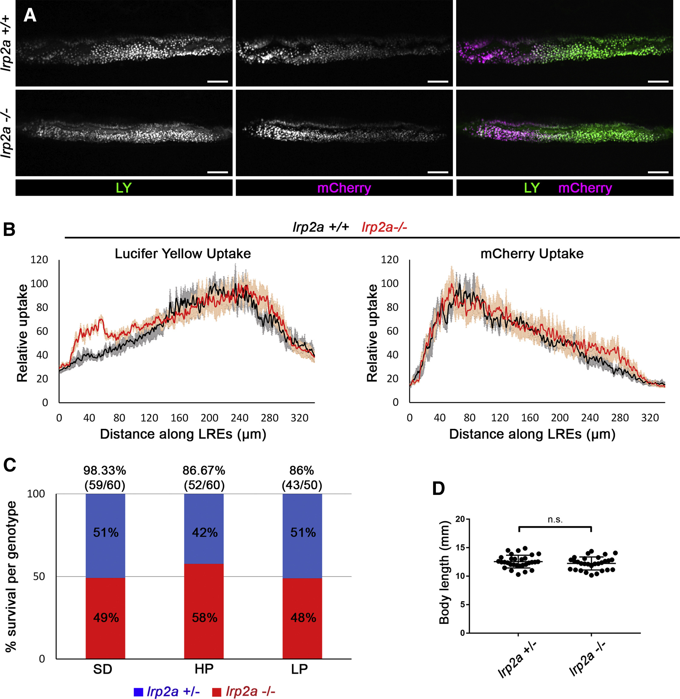
lrp2a (megalin) Mutants Do Not Show Defects in LRE Uptake or Survival under Different Feeding Conditions
(A) Live confocal images showing LY or mCherry uptake in LREs of lrp2amw1 +/+, −⁄− 6 dpf larvae. The larvae were imaged 5 h after gavage. Scale bar, 20 μm.
(B) Internalization profiles of LY (left) and mCherry (right) along LREs of 6 dpf lrp2amw1 +/+ (n = 5), −⁄− (n = 5). Data are means ± S.E.M.
(C) Survival rates of lrp2amw1 +/− and −⁄− 30 dpf larvae raised under different feeding conditions: non-calorie restricted standard diet (SD), calorie-restricted high-protein diet (HP), and calorie-restricted low-protein diet (LP). Larvae from the cross lrp2amw1 +/− X lrp2amw1 −⁄−were used for the experiment. At 30 dpf, the survived larvae were counted and genotyped to determine what percentage of survived larvae are lrp2a +/− or −⁄−. The numbers above the graph bars indicate the percentage of overall tank survival for each feeding condition.
(D) Comparison of body lengths of lrp2amw1 +/− (n = 30) and −⁄− (n = 29) 30 dpf larvae raised under non-calorie restricted SD. Two-tailed unpaired t test was used for statistical analysis. p values, t values and degree of freedom for the statistical tests are provided in the Table S3.
Reprinted from Developmental Cell, 51(1), Park, J., Levic, D.S., Sumigray, K.D., Bagwell, J., Eroglu, O., Block, C.L., Eroglu, C., Barry, R., Lickwar, C.R., Rawls, J.F., Watts, S.A., Lechler, T., Bagnat, M., Lysosome-Rich Enterocytes Mediate Protein Absorption in the Vertebrate Gut, 7-20.e6, Copyright (2019) with permission from Elsevier. Full text @ Dev. Cell