IMAGE

Figure 1

ID
ZDB-IMAGE-191230-958
Source
Figures for Kobayashi et al., 2019
Image
Figure Caption

Figure 1

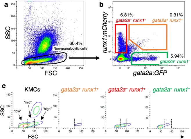
Isolation of HSPCs from the zebrafish kidney. FCM analysis of KMCs was performed in a gata2a:GFP; runx1:mCherry double-transgenic zebrafish. (a) A scatter profile of KMCs. The SSClow non-granulocytic cell fraction is gated. (b) SSClow cells are subdivided into three distinct hematopoietic populations, gata2a+runx1+ (orange gate), gata2arunx1+ (red gate), and gata2a+runx1 (green gate). (c) Contour plot of FSC vs. SSC in KMCs, gata2a+runx1+, gata2arunx1+, and gata2a+runx1 cells. Dotted lines separate the “low”, “mid”, and “high” intensity of FSC.

Acknowledgments
This image is the copyrighted work of the attributed author or publisher, and ZFIN has permission only to display this image to its users. Additional permissions should be obtained from the applicable author or publisher of the image. Full text @ Sci. Rep.