IMAGE

Fig. 5

ID
ZDB-IMAGE-191230-693
Source
Figures for Pérez-Rius et al., 2019
Image
Figure Caption

Fig. 5

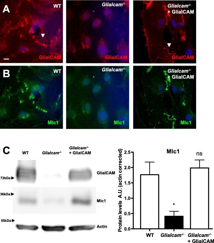
Mlc1 is mislocalized in primary Glialcam−/− astrocytes. Localization of GlialCAM (a) and Mlc1 (b) in primary astrocytes from wild-type (WT, left), Glialcam−/− (middle) and Glialcam−/− complemented with adenoviruses expressing human GlialCAM (right). In WT and complemented astrocytes, GlialCAM and Mlc1 are located at cell-cell junctions (arrowheads). Scale bar: 10 μm. (c) GlialCAM and Mlc1 protein levels primary astrocytes from wild-type (WT, left), Glialcam−/− (middle) and Glialcam−/− complemented with an adenoviruss expressing human GlialCAM. Actin served as a loading control. Two other independent experiments gave similar results. Densitometric analysis (n = 3) indicates that Mlc1 levels were reduced in astrocytes from Glialcam−/− mice and expression was recovered after expression of GlialCAM using adenoviruses. * p < 0.05 vs wild-type astrocytes

Acknowledgments
This image is the copyrighted work of the attributed author or publisher, and ZFIN has permission only to display this image to its users. Additional permissions should be obtained from the applicable author or publisher of the image. Full text @ Orphanet J Rare Dis