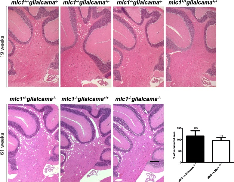
IMAGE

Fig. 3

ID
ZDB-IMAGE-191230-690
Source
Figures for Pérez-Rius et al., 2019
Image
Figure Caption

Fig. 3

Myelin vacuolization in Glialcam−/−, Mlc1−/− and Glialcam−/−Mlc1−/− mouse models. Haematoxylin-eosin staining of sagittal sections of the cerebellum of 19- and 61-week-old mice showed similar levels of myelin vacuolization in Glialcam−/−, Mlc1−/− and Glialcam−/−Mlc1−/− animals. As a control we show the same area of a wild-type mouse at 19 weeks. The inset shows the percentage of vacuolation in double KO animal versus the vacuolation observed in Glialcam KO animals (n = 3) and Mlc1 KO animals (n = 3) considering each age independently, without substracting the minor vacuolization observed in wild-type animals. Data were analyzed by GraphPad Prism software. In order to compare the different groups (dKO vs Glialcam−/− and dKO vs Mlc1−/−), one-way analysis of variance (ANOVA) followed by post-hoc Bonferroni’s multiple comparison test was used. ns: not significative. Scale bar, 400 μm

Acknowledgments
This image is the copyrighted work of the attributed author or publisher, and ZFIN has permission only to display this image to its users. Additional permissions should be obtained from the applicable author or publisher of the image. Full text @ Orphanet J Rare Dis