IMAGE

Fig. 2

ID
ZDB-IMAGE-191230-689
Source
Figures for Pérez-Rius et al., 2019
Image
Figure Caption

Fig. 2

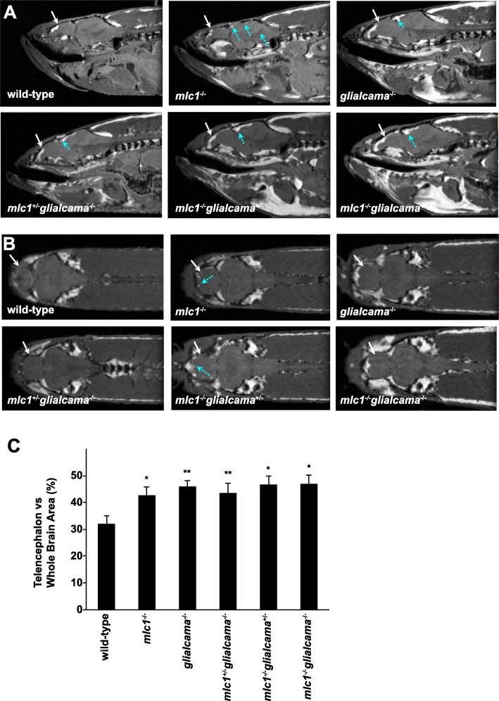
MR images of wild-type and various zebrafish mutants. a The sagittal slices were obtained with an in-plane resolution of 47 μm. The size of telencephalon was bigger in mutant zebrafish as compared to wild type (white arrows). Lesions in mesencephalon in mutants are shown with blue arrows. b The coronal slices were obtained with an in-plane resolution of 47 μm. The size of telencephalon was bigger in mutant zebrafish as compared to wild type (white arrows). Lesions in telencephalon are shown with blue arrows. c Telencephalon vs. whole brain area (%). In order to compare the size of the telencephalon relative to the whole brain of the different groups one-way analysis of variance (ANOVA) was performed and indicated that the size was larger in all mutants as compared to wild type (*p < 0.05; **p < 0.005) (n = 3). No statistical differences were observed in the percent area of telencephalon versus whole brain size between single knockout zebrafish for one gene with the single knockout/heterozygous or the double knockout (p > 0.05) (see Additional file 3: Table S1)

Figure Data
Acknowledgments
This image is the copyrighted work of the attributed author or publisher, and ZFIN has permission only to display this image to its users. Additional permissions should be obtained from the applicable author or publisher of the image. Full text @ Orphanet J Rare Dis