IMAGE

Fig. 1

ID
ZDB-IMAGE-191230-45
Genes
Source
Figures for Hu et al., 2019
Image
Figure Caption

Fig. 1

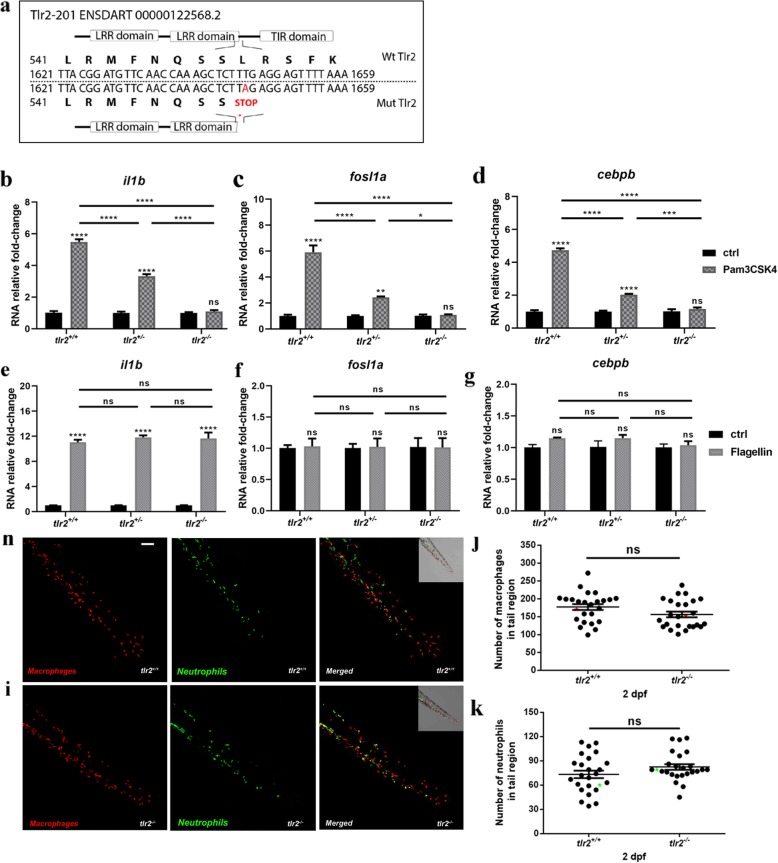
Characterization of the Tlr2 mutant. a mutant DNA and protein sequence. A point mutation (T to A) in the C-terminal of the second LRR domain of zebrafish Tlr2 introduces a premature stop codon. The predicted truncated protein lacks the whole TIR domain. Nucleotide and amino acid positions are indicated with respect to the translation start codon. tlr2+/+, tlr2+/− and tlr2−/− embryos were injected at 27 hpf with 1 ng Pam3CSK4 (b-d) or 0.1 ng flagellin (e-g) and expression levels of il1b, fosl1a and cebpb were determined at 1 h post injection by qPCR. Data (mean ± SEM) are combined from three biological replicates (n = 10 embryos per group) and expressed relative to their corresponding mock injection (water) control (ctrl), which is set at 1. Statistical significance of differences between ctrl and PAMPs injection groups was determined by two-way ANOVA with Tukey’s Multiple Comparison method as a post-hoc test (b-g), ns, non-significant; *, P < 0.05; ***, P < 0.001; ****, P < 0.0001. h, i. Representative Stereo images of the caudal hematopoietic tissue of tlr2+/+Tg (mpeg1:mCherry-F);TgBAC (mpx: EGFP) (h) and tlr2−/−Tg (mpeg1:mCherry-F);TgBAC (mpx: EGFP) (i) were taken at 2 dpf for quantification of macrophages and neutrophils numbers. At 2 dpf, numbers of mCherry-labeled macrophages (j) and GFP-labeled neutrophils (k) were counted using Leica TCS SP8 confocal laser scanning microscopy (CLSM) of transgenic lines. Data (mean ± SEM) were combined from two independent experiments. No significant differences (ns) in the number of macrophages (j) and neutrophils (k) was detected with a t-test

Figure Data
Acknowledgments
This image is the copyrighted work of the attributed author or publisher, and ZFIN has permission only to display this image to its users. Additional permissions should be obtained from the applicable author or publisher of the image. Full text @ BMC Genomics