IMAGE

Figure 4

ID
ZDB-IMAGE-191230-1901
Source
Figures for Xu et al., 2019
Image
Figure Caption

Figure 4

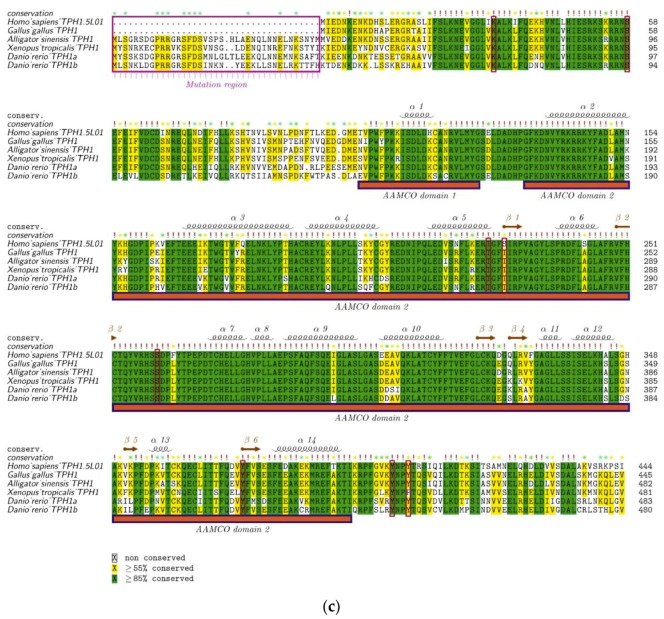
Alignment and secondary structures of TPH protein sequences in vertebrates. (a) TPH2 protein sequences from representative vertebrate species were aligned with the human TPH2 and its secondary structure template 4V06. (b) Sequence alignment between TPH1a and TPH1b in some representative teleosts is provided for comparison. (c) TPH1 protein sequences from representative vertebrates were aligned with the human TPH1 and its secondary structure template 5L01. The mutation regions are marked with rose boxes and upper arrows in each figure. The red boxes denote the phosphorylation sites based on the human templates. Related alignment and secondary structures in more vertebrate species are provided in Figure S2.

Acknowledgments
This image is the copyrighted work of the attributed author or publisher, and ZFIN has permission only to display this image to its users. Additional permissions should be obtained from the applicable author or publisher of the image. Full text @ Genes (Basel)