IMAGE

Figure 1

ID
ZDB-IMAGE-191230-1635
Source
Figures for Yang et al., 2019
Image
Figure Caption

Figure 1

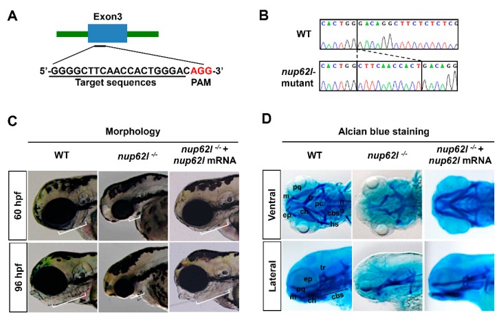
Loss of Nup62l led to severely impaired formation of PA cartilages. (A) Diagram showing the CRISRP/Cas9 target DNA sequence (underline) of zebrafish gene nup62l. PAM region (AGG) was shown in red. (B) Sanger sequencing results revealed an 11-bp genomic DNA fragment insertion at the target site in nup62l-mutants. (C) Lateral views showing the pharyngeal morphology of WT siblings. Embryos of nup62l-mutants at 60 hpf or 96 hpf were injected with or without 300 pg nup62l mRNA/embryo. White lines indicated the pharyngeal regions. Note a shortened and thinned pharyngeal region in nup62l-mutants. (D) Images showing pharyngeal cartilage elements stained with alcian blue in WT. The nup62l-mutants injected with or without 300 pg nup62l mRNA at 96 hpf were ventrally viewed in upper panels and laterally viewed in lower panels. cbs, ceratobranchials; ch, ceratohyal; ep, ethmoid plate; hs, hyosymplectic; m, Meckel’s cartilage; no, notochord; pc, parachordal; pq, palatoquadrate; tr, trabecula.

Figure Data
Acknowledgments
This image is the copyrighted work of the attributed author or publisher, and ZFIN has permission only to display this image to its users. Additional permissions should be obtained from the applicable author or publisher of the image. Full text @ Cells