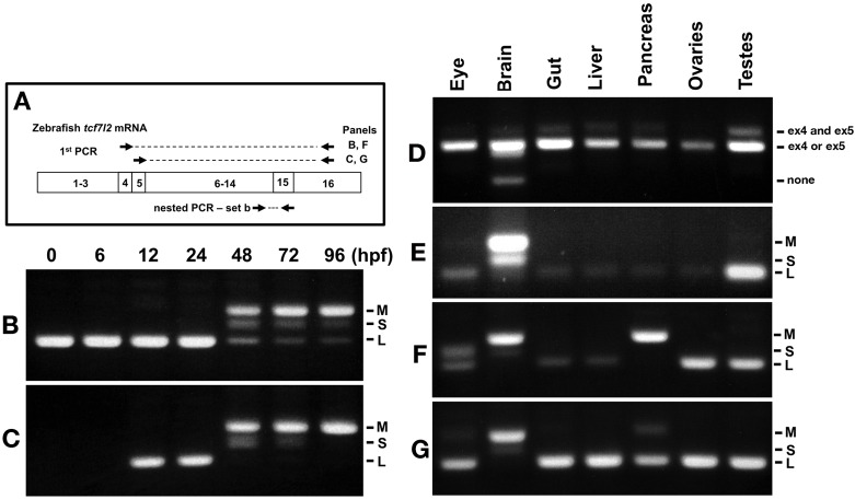
IMAGE

Figure 2.

ID
ZDB-IMAGE-191230-1595
Genes
Source
Figures for Young et al., 2019
Image
Figure Caption

Figure 2. Expression of <italic>tcf7l2</italic> alternative exons 4, 5 and 15 varies across development and in adult organs.

RT-PCR analysis of alternative exons 4, 5 and 15 of zebrafish tcf7l2 across development and in various adult organs. (A) Schematic representation of nested RT-PCR strategy used for panels B, C, F and G. (B–C) cDNA from embryos of ages indicated (hpf) was PCR amplified using a forward primer that anneals over exon 4 (B) or exon 5 (C) and a reverse primer that anneals to exon 16 common to all tcf7l2 mRNA variants. The product from this first PCR was then used as a template for a nested PCR using primer set ‘b’ (as in Figure 1A) that amplifies exon 15 and reveals the different possible Ct ends of Tcf7l2. This last PCR product is shown in these panels. M (Medium), S (short) and L (Long) C-terminal Tcf7l2 variant end. (D–E) RT-PCR experiments performed on cDNA of the indicated adult organs using primer set ‘a’ (Materials and methods) amplifying the region of alternative exons 4 and 5 (D) or using primer set ‘b’ (Materials and methods) amplifying the region of alternative exon 15 (E). (F–G) Same PCR amplification strategy used in panels (B–C) to detect the C-terminal Tcf7l2 variants associated with exons 4 or 5, but using the indicated adult organ cDNA as template in the 1st PCR reaction.

Figure Data
Acknowledgments
This image is the copyrighted work of the attributed author or publisher, and ZFIN has permission only to display this image to its users. Additional permissions should be obtained from the applicable author or publisher of the image. Full text @ Elife