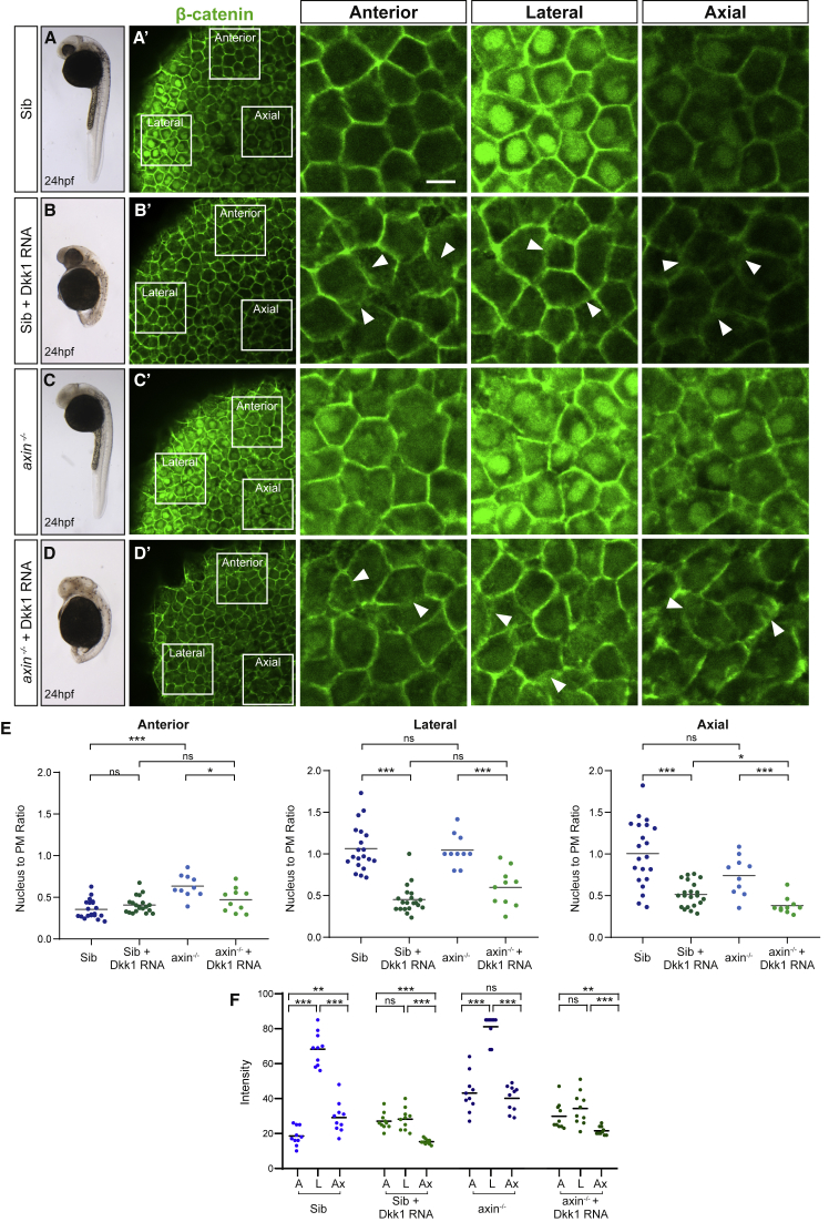
IMAGE

Figure 7

ID
ZDB-IMAGE-191230-1580
Source
Figures for Johansson et al., 2019
Image
Figure Caption

Figure 7

Dkk1 Sequesters β-Catenin at the Plasma Membrane

(A–D) Sibling and axin1 mutant embryos at 24 h post-fertilization (hpf) (left column) and 80% epiboly (EB) with (B) and (D) or without (A) and (C) Dkk1 misexpression. 80% EB embryos were stained with an antibody against β-catenin (green) to assess endogenous β-catenin distribution in different subcellular compartments in various regions of the embryo. White-bordered squares in the second column indicate the location of single-plane confocal images in three regions of the embryo shown at a higher magnification in the three last columns. Scale bar, 20 μm. (A) Sibling embryos show patterned distribution of β-catenin (green) in different regions of the embryo at tissue and subcellular levels. In the anterior region β-catenin is detected exclusively at the plasma membrane, while high levels are seen in both nuclei and the plasma membrane, laterally. Axial cells show a low level of β-catenin in both nuclei and plasma membrane. (B) Dkk1-expressing sibling embryos display large heads and extension defects at 24 hpf and show a loss of nuclear β-catenin (green) in anterior, lateral, and axial regions of the embryo but maintain β-catenin at the plasma membrane despite the loss of cell-cell adhesion. Arrowheads point to diffuse membrane morphology. (C) axin1 null mutants show a loss of anterior fate at 24 hpf and β-catenin-positive (green) nuclei in the anterior region at 80% EB. Contrary to the current model, these gastrula embryos display the same relative subcellular distribution of β-catenin found in normal siblings across the three areas measured. (D) Despite the expected lack of telencephalon and eyes, the Dkk1-expressing axin1 mutants show an overall AP organization similar to Dkk1-expressing embryos at 24 hpf. Similar to Dkk1-RNA-injected siblings, β-catenin was retained at the plasma membrane, and cells displayed a loss of cell-cell adhesion. Arrowheads point to diffuse membrane morphology.

(E) Nucleus to plasma membrane (PM) β-catenin (green) fluorescence intensity ratios quantified in anterior, lateral, and axial cells in sibling (Sib) and axin1 mutant (axin−/−) embryos, with or without Dkk1 expression. A significantly higher proportion of β-catenin is present at the PM in Dkk1-expressing Sibs and mutants in the lateral and axial regions than control embryos. Fluorescence intensity ratios were measured in 10 cells per region in each embryo (Sib, n = 2; Sib + Dkk1 RNA, n = 2; axin−/−, n = 1; and axin−/− + Dkk1 RNA, n = 1). p > 0.05 ns,p < 0.05, and ∗∗∗p < 0.001.

(F) Absolute levels of β-catenin nuclear expression, calculated by fluorescence intensity in 10 cells in each of the three regions per embryo. A, anterior; L, lateral; and Ax, axial. p > 0.05 ns, ∗∗p < 0.01, ∗∗∗p < 0.001.

Acknowledgments
This image is the copyrighted work of the attributed author or publisher, and ZFIN has permission only to display this image to its users. Additional permissions should be obtained from the applicable author or publisher of the image.

Reprinted from Developmental Cell, 51(6), Johansson, M., Giger, F.A., Fielding, T., Houart, C., Dkk1 Controls Cell-Cell Interaction through Regulation of Non-nuclear β-Catenin Pools, 775-786.e3, Copyright (2019) with permission from Elsevier. Full text @ Dev. Cell