IMAGE

Fig. 2

ID
ZDB-IMAGE-191230-1489
Source
Figures for Hamada et al., 2019
Image
Figure Caption

Fig. 2

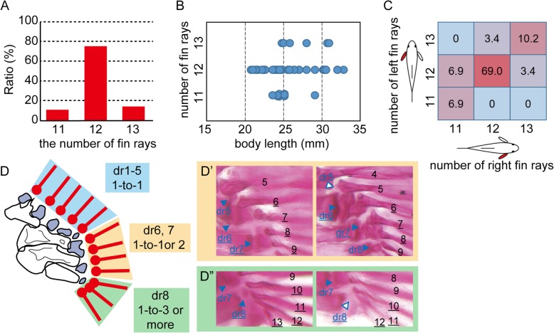
Observation of skeletal anatomy of the pectoral fin in the RW strain. a Distribution of the variation in fin ray number. b Relationship between the number of rays and body length. Blue circles indicate individuals. c Ratio of the combination of the numbers of fin rays in the left and right pectoral fins. d. Schematic drawing of the connection between the fin rays (red bar with circle) and distal radials (dark blue). Light blue, orange, and green boxes indicate the relationship in the one-to-one, one-to-one or two, and one-to-three or more modes, respectively. d’. Connection between fin rays and the sixth and seventh radials. The seventh radial showed connection in a one-to-two mode; the sixth radial showed the connection in a one-to-two (left panel) or a one-to-one (right panel) mode. d”. Connection between the fin rays and the eighth radial. The number of fin rays connected to the eighth radial varied (four rays in the left panel and three rays in the right panel). Blue-colored arrowheads with dr5–8 indicate the 5th to 8th distal radials, respectively. Blue-outlined arrowheads with dr5 in the right panel of d’ and dr8 in the right panel of d” indicate the absence of the 5th and 8th distal radials, respectively. Black-colored numbers indicate fin ray numbers. Underlined characters in d’ indicate the 6th and 7th distal radials and the fin rays connected to them. Underlined characters in d” indicate the 8th distal radial and the fin rays connected to it

Acknowledgments
This image is the copyrighted work of the attributed author or publisher, and ZFIN has permission only to display this image to its users. Additional permissions should be obtained from the applicable author or publisher of the image. Full text @ Zoological Lett