IMAGE

Fig. 4

ID
ZDB-IMAGE-191230-1444
Source
Figures for Carter et al., 2019
Image
Figure Caption

Fig. 4 Inhibition of both MCL-1 and BCL-X<sub>L</sub> induces apoptosis in SCCHN cell lines and primary cells.

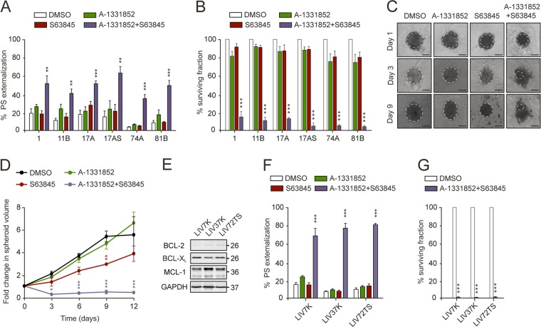
SCCHN cell lines exposed to a combination of A-1331852 and S63845 (100 nM each) for 24 h exhibited a enhanced apoptosis and b decreased clonogenicity, compared to the individual BH3 mimetics. c Phase contrast images (scale bars 100 μm), and d line graph to show fold change in spheroid volume over 12 days, following exposure to the specified drug(s). Dotted lines in c demarcate the outline of each intact spheroid. e Western blot analysis of antiapoptotic protein levels in SCCHN primary cells. These cells, exposed to a combination of A-1331852 and S63845 (100 nM) for 4 h exhibited f enhanced apoptosis and g decreased clonogenicity, compared to the indicated treatments. a, b, f One-way ANOVAs with Dunnett’s multiple comparisons tests, with a single pooled variance; d two-way repeated measures ANOVA with Dunnett’s multiple comparisons test; g unpaired, two-tailed t tests. Error bars = mean ± SEM of at least three independent experiments. *P < 0.05, **P < 0.01, ***P < 0.001.

Acknowledgments
This image is the copyrighted work of the attributed author or publisher, and ZFIN has permission only to display this image to its users. Additional permissions should be obtained from the applicable author or publisher of the image. Full text @ Cell Death Dis.