
Image
|
Figure Caption
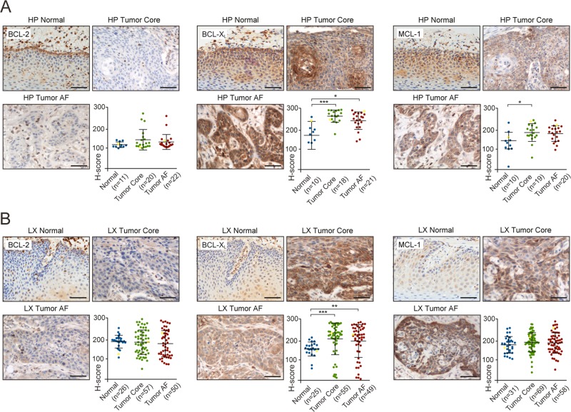
Fig. 2
Representative IHC images of normal tissue, primary tumor core and advancing front (AF) from
Acknowledgments
This image is the copyrighted work of the attributed author or publisher, and
ZFIN has permission only to display this image to its users.
Additional permissions should be obtained from the applicable author or publisher of the image.
Full text @ Cell Death Dis.