IMAGE

Fig 2

ID
ZDB-IMAGE-191230-1363
Source
Figures for Pazhakh et al., 2019
Image
Figure Caption

Fig 2 Variant shuttles of fungal conidia from neutrophil to macrophage.

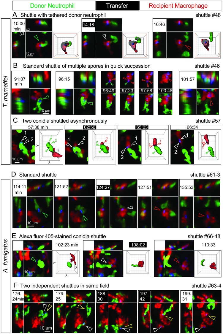
A variety of shuttles of conidia (blue) from Tg(mpx:EGFP) neutrophils (green) to Tg(mpeg1:Gal4FF)×(UAS-E1b:Eco.NfsB-mCherry) macrophages (red). In each example, panels include isometric orthogonal yz and xz views corresponding to the xy maximal intensity projection and indicate the time in min from start of movie. Time points colored white-on-black are the moments of transfer. (A,C,E) include volume-rendered views corresponding to the maximal intensity projection; where this volume is sectioned (as in panel A), the framing box is shown in red. Colored arrowheads indicate the conidium within donor neutrophil (green), at the point of intercellular transfer (white), and in the recipient macrophage (red). (A–C) Shuttles of T. marneffei conidia. (A) Shuttle demonstrating tethering of the donor neutrophil at the moment of transfer. (B) Shuttle of multiple conidia from one donor neutrophil in quick succession. First two frames show donor neutrophil laden with multiple conidia at two preshuttle time points. Frames from t = 96:49–100:48 min are maximal intensity projections only, encompassing the shuttling transfer of 3 conidia over 4 min Upper row of panels shows red (macrophage) and blue (conidia) channels only; lower row of panels includes the green channel (donor neutrophil). (C) Shuttle of 2 conidia from one donor neutrophil one after the other at an interval of 2 min 13 s. Volume-rendered images corresponding to maximal intensity projections show the 2 shuttled conidia (labeled 1 and 2) before, during, and after the shuttle. (D–F) Shuttles of A. fumigatus conidia. (D) Standard shuttle of single conidium. (E) Standard shuttle of conidium labeled with Alexa Fluor 405 rather than calcofluor, accompanied by volume-rendered images that focus attention onto the conidium and donor neutrophil of interest. (F) Two independent shuttles by different donor neutrophils occurring in the same field. In this series, the course of each shuttled spore is followed by white and yellow arrowheads. Scales as shown. Stills in A–F correspond to S1C, S1E, S1F, S2A, S2C and S2E Movies, respectively. Eco.Nfsb, E. coli nitroreductase; EGFP, enhanced green fluorescent protein; Gal4FF, engineered form of S. cerevisiae Gal4 transcriptional activator; mpeg1, macrophage-expressed gene 1; mpx, myeloid-specific peroxidase; Tg, transgenic; UAS-E1b, upstream activating sequence fused to minimal adenovirus E1b promoter.

Acknowledgments
This image is the copyrighted work of the attributed author or publisher, and ZFIN has permission only to display this image to its users. Additional permissions should be obtained from the applicable author or publisher of the image. Full text @ PLoS Biol.