IMAGE

Figure 3

ID
ZDB-IMAGE-191230-1290
Genes
Source
Figures for Chen et al., 2019
Image
Figure Caption

Figure 3

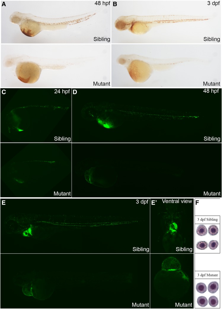
Deficiency in rps9 induces anemia in mutant embryos. (A-B) Detection of hemoglobin levels by o-dianisidine staining for rps9 in both siblings and mutants at 48 hpf and 3 dpf. (C-E, E’) Detection of erythroid cells by crossing rps9 mutant into LCR:EGFP transgenic fish line. At 24 hpf, the number of blood cells are reduced slightly; At 48 hpf and 3 dpf, erythrocytes are almost deprived in the trunk, and only a small amount of red blood cells remain in the cardiac region. (F) Wright-Giemsa staining of erythroid cells at 3 dpf in sibling and mutant.

Figure Data
Acknowledgments
This image is the copyrighted work of the attributed author or publisher, and ZFIN has permission only to display this image to its users. Additional permissions should be obtained from the applicable author or publisher of the image. Full text @ G3 (Bethesda)