IMAGE

Figure 3

ID
ZDB-IMAGE-191230-1206
Source
Figures for Kefalos et al., 2019
Image
Figure Caption

Figure 3

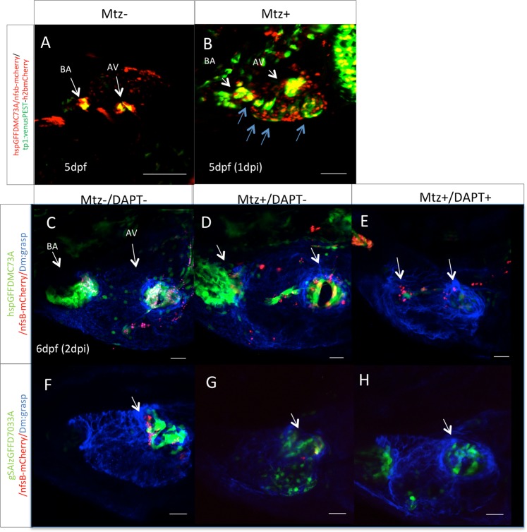
Notch signaling is activated during valve regeneration. (A) Confocal z-stack of a Tg(TP1:VenusPEST)/hspGFFDMC73A: UAS-E1b:NfsB-mCherry double transgenic embryo at 5 dpf. Tg(TP1:VenusPEST) signal of activated Notch signaling is restricted to the valves. Scale bar: 100 microns. (B) 1 day post MTZ treatments. Expansion of activation of Notch signaling throughout the ventricle is observed (blue arrows) Scale bar: 50microns. Confocal z-stacks of untreated Tg(hspGFFDMC73A: UAS-E1b:NfsB-mCherry) (C) and Tg(GSAIzGFFD703A: UAS-E1b:NfsB-mCherry) (F) double transgenic embryos at 6 dpf. Confocal z-stack of MTZ treated Tg(hspGFFDMC73A: UAS-E1b:NfsB-mCherry) (D), and Tg(GSAIzGFFD703A: UAS-E1b:NfsB-mCherry) (G), at 6 dpf. Confocal z-stack of MTZ treated and then during regeneration 48 hours DAPT treated Tg(hspGFFDMC73A: UAS-E1b:NfsB-mCherry) (E), and Tg(GSAIzGFFD703A: UAS-E1b:NfsB-mCherry) (H), at 6 dpf. (I) Each experiment was carried out at least 3 independent times with n = 10 embryos per experimental group. Scale bars: 20 micron.

Acknowledgments
This image is the copyrighted work of the attributed author or publisher, and ZFIN has permission only to display this image to its users. Additional permissions should be obtained from the applicable author or publisher of the image. Full text @ Sci. Rep.