IMAGE

Figure 2

ID
ZDB-IMAGE-191230-1205
Source
Figures for Kefalos et al., 2019
Image
Figure Caption

Figure 2

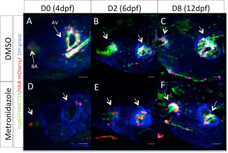
Zebrafish embryonic valves can regenerate. (A–C) Confocal z-stacks of untreated Tg(hspGFFDMC73A/UAS-E1b:NfsB-mCherry) larvae at 4 dpf, 6 dpf and 12 dpf respectively. (D–F) Confocal z-stacks of MTZ treated Tg(hspGFFDMC73A/UAS-E1b:NfsB-mCherry) and larvae at 4 dpf, 6 dpf and 12 dpf respectively. Arrows show the ablation of mCherry+ cells at the AV canal and OFT at 4 dpf. From 6 dpf more mCherry+ cells could be observed at the AV canal. By 12 dpf the regeneration process could be imaged, as mCherry+, AV differentiated cells were present at both valves. Scale bars: 20 microns. Each experiment was carried out at least 3 independent times with n = 10 embryos per experimental group.

Acknowledgments
This image is the copyrighted work of the attributed author or publisher, and ZFIN has permission only to display this image to its users. Additional permissions should be obtained from the applicable author or publisher of the image. Full text @ Sci. Rep.