IMAGE

Fig 6

ID
ZDB-IMAGE-191230-1178
Source
Figures for Lischik et al., 2019
Image
Figure Caption

Fig 6 The established toolbox enables imaging of previously challenging organs and tissues, such as the retina and the brain.

Using the pigment double knockout in combination with α-Bungarotoxin, eGFP and mCherry coupled to histone2a the imaging was conducted in a single plane illumination microscope (SPIM) we could image the head region of wild-type and
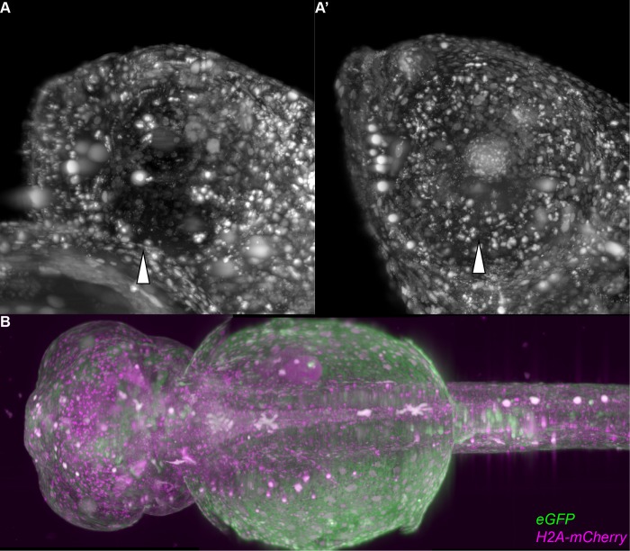
 spooky embryos. Hollow arrowheads point towards the eyes in A and B. (A) Maximum projection of H2A-mCherry signal of a lateral view of a wild-type medaka embryo. The eye is not penetrable and no nuclei are visible within or behind it. (A') Maximum projection of H2A-mCherry signal of a lateral view of a spooky medaka embryo. The eye is penetrable and nuclei are visible within or behind it. (B) Stitched maximum projections of a dorsal view of a spooky embryo. This embryo was imaged for 48 hours, which can be seen in S3 Movie.

Acknowledgments
This image is the copyrighted work of the attributed author or publisher, and ZFIN has permission only to display this image to its users. Additional permissions should be obtained from the applicable author or publisher of the image. Full text @ PLoS One