IMAGE

Figure 6

ID
ZDB-IMAGE-191230-1033
Source
Figures for Griffin et al., 2019
Image
Figure Caption

Figure 6

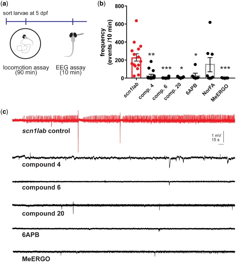
Electrophysiological assay to identify drugs that rescue the scn1lab mutant epilepsy phenotype. (a) Electrophysiology recording were obtained with an electrode placed in the forebrain of 5 dpf agar-immobilized scn1lab larvae that had previously showed suppressed seizure-like behaviour in the locomotion assay. (b) Bar graphs show the frequency of epileptiform events in a 10 min recording epoch for scn1lab larvae exposed to clemizole analogues 4 (n = 15), 6 (n = 12), 20 (n = 9), 6-APB (n = 6), norfenfluramine (NorFA) (n = 8), methylergonovine (MeERGO) (n = 7) or scn1lab mutants (n = 15). The graph represents mean ± SEM and individual data points are shown. Kruskal–Wallis one-way analysis of variance was used to test for significance. *P <0.05; **P <0.01; ***P <0.001. (c) Representative field electrode recording epochs (10 min) are shown for clemizole analogues 4, 6, 20, methylergonovine (MeERGO) and 6-APB. These compounds showed significant changes in the frequency of events compared to untreated scn1lab mutant zebrafish (red).

Acknowledgments
This image is the copyrighted work of the attributed author or publisher, and ZFIN has permission only to display this image to its users. Additional permissions should be obtained from the applicable author or publisher of the image. Full text @ Brain Commun