
|
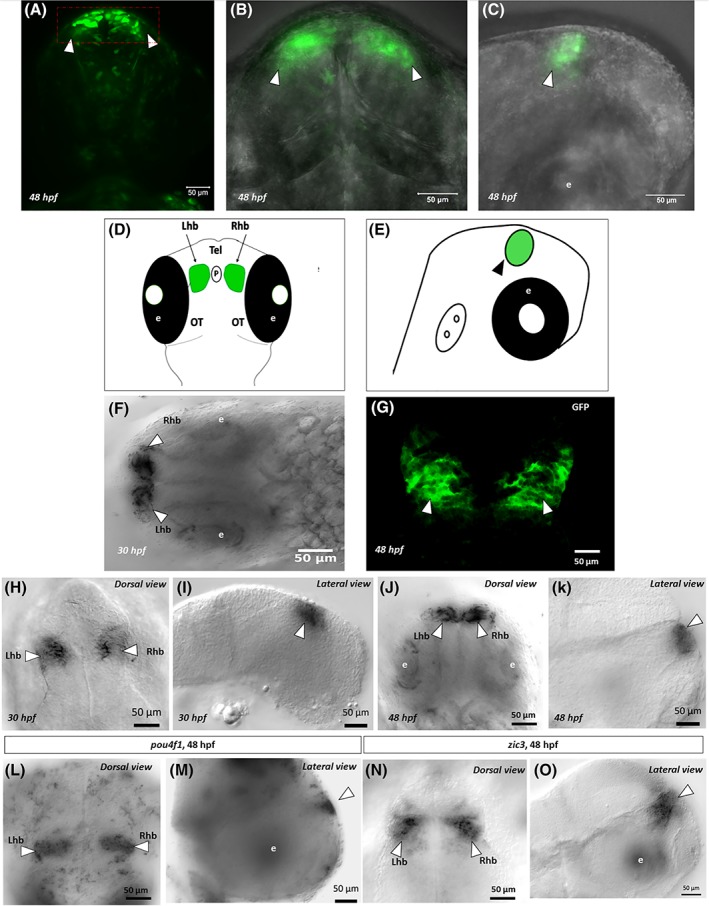
Figure 3
Green fluorescent protein expression in stable F1 line at 48 hpf. A, Green fluorescent protein expression in live embryos is indicated by arrowheads in the left and right habenula. B, The same GFP expression from the same embryo in a closer view. C, Green fluorescent protein expression from the lateral side. D and E, Schematics that show GFP expression in the habenula at 48 hpf in the dorsal and lateral views. F, RNA whole‐mount in situ hybridization using a probe against GFP, confirming its expression in the habenula at 30 hpf. G, Whole‐mount anti‐GFP immunohistochemistry, confirming GFP expression in the habenula at 48 hpf. H‐K, RNA whole‐mount in situ hybridization against GFP, showing GFP expression in the habenula at 30 hpf (H, I) and 48 hpf (J, K). L and M, RNA whole‐mount in situ hybridization using