
|
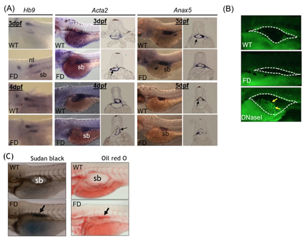
Fig. 2
The tissue integrity and pathology of the swim bladder in wild-type and FD embryos and larvae. Heat-shocked larvae of both wild-type control and Tg( hsp:EGFP-GGH) were harvested at indicated stages post embryogenesis and analyzed for swim bladder development. ( A) Embryos were subjected to WISH with tissues specific probes including Hb9 (epithelial layer), Acta2 (mesenchyme layer, smooth muscle) and Anax5 (outer mesothelium layer). Cross-sections of the embryos after WISH staining revealed comparable intensity and morphology for the tissue layers composed of swim bladders (black arrows) in the early embryos. ( B) Larvae at 5 dpf examined with TUNEL assay for apoptotic cells (arrows) revealed no positive signal in the swim bladder area (circled by dash-line) of both wild-type control and FD larvae. Embryos pre-treated with DNase I served as a positive control. ( C) Larvae at 5 dpf were stained with Sudan black (left) and Oil red O (right) to reveal outlines of swim bladder (black arrows) in larvae. nt, neural tube; sb, swim bladder; WT, wild-type larvae with heat-shock; FD, FD larvae with heat-shock.