
|
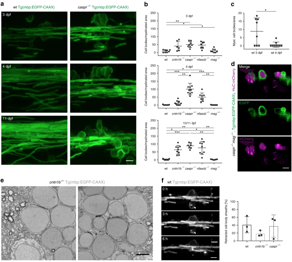
Fig. 1
Loss of adhesion molecules of the paranodal axo-glial junction results in neuronal cell body wrapping. a CNS myelin of 3, 4 and 10 dpf wild-type (wt) and caspr−/− fish. Note cell body wrappings in caspr−/− fish. b Cell body wrappings per myelinated area at 3, 4, and 10/11 dpf ( n = 6–10, Kruskal Wallis ANOVA: 3 dpf: p = 0.0004, 4 dpf and 10/11 dpf: p < 0.0001). c Cell body wrappings in wt fish ( n = 8–9). Data derived from panel ( b). Mann-Whitney test, * p = 0.0399. d HuC:mCherry expression in 4 dpf caspr−/− mag−/− fish shows neuronal cell bodies enwrapped by myelin membrane. e SEM images of cell body wrappings in cntn1b−/− fish at dpf 10. f Sheath retractions (arrow) from neuronal cell bodies (30 min intervals, 15–16 h time lapse experiments, n = 3, Kruskal-Wallis one-way ANOVA: p = 0.4393). Images ( a, d, f) are maximum intensity projections of Tg(mbp:EGFP-CAAX) zebrafish dorsal spinal cord. p values, * < 0.05, ** < 0.01, *** < 0.001. Data are presented as means ± s.d.Scale bars, 5 mm ( a, d, f), 2 µm ( e). Source data are provided as a Source Data file