
|
Fig. S7
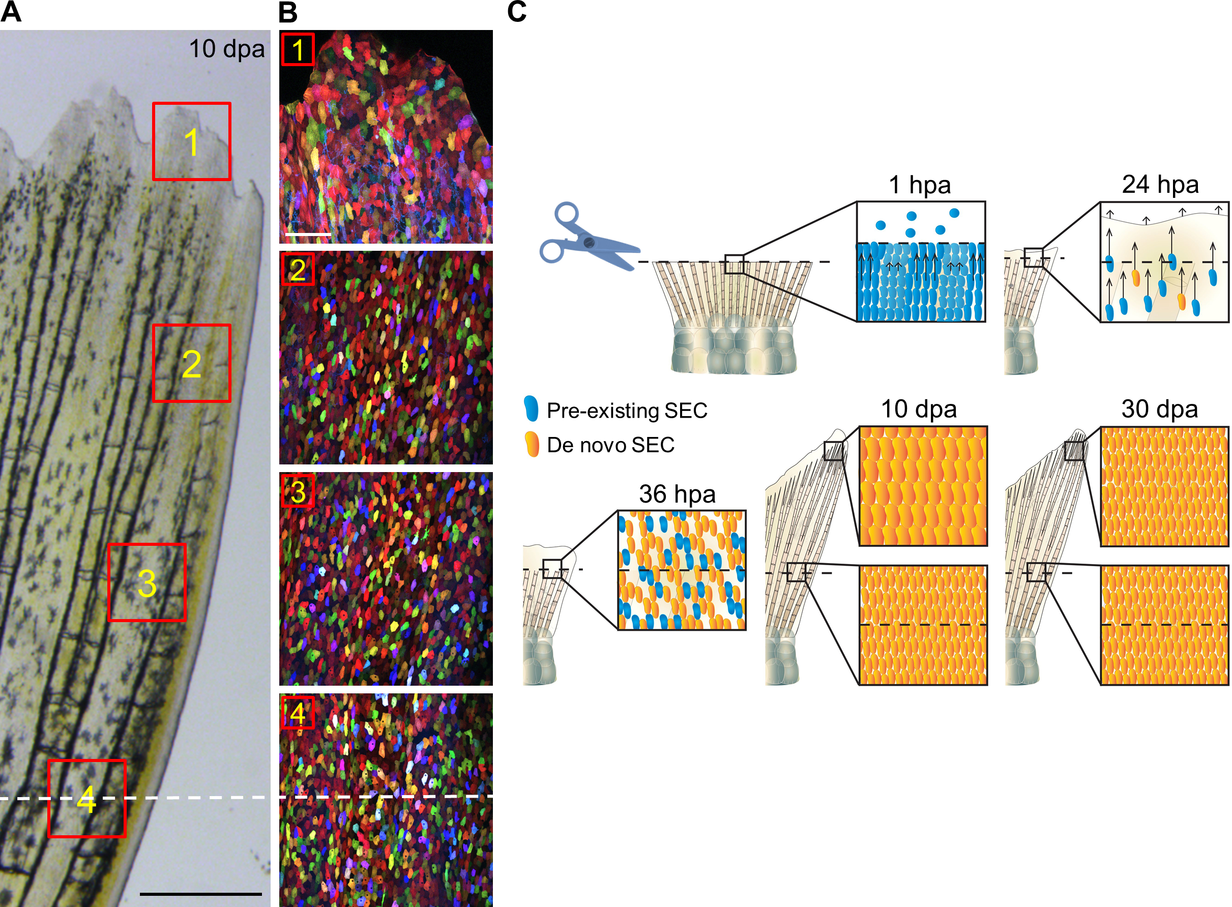
Sequence of Collective SEC Behaviors During Fin Regeneration, Related to Figure 6. (A, B)
Brightfield view of adult zebrafish caudal fin at 10 dpa. Scale bars, 0.5 mm. Red boxes in (A) indicate areas where
z-stacked confocal images (B) were captured. Scale bars, 100 μm. White-dashed lines indicate the amputation plane.
Images in (A) and (B) were captured from the same animal. Scale bars, 100 μm. (C) Wounds seal in the first hour
post amputation (hpa) by interactions between alternating SEC sheets with distinct cell displacement and shape
responses. Many SECs are extruded at the amputation plane during this process. At 24 hpa, spared SECs are
recruiting to the regenerating area over long distances. By 36 hpa, do novo SECs are well-integrated with preexisting
SECs, and new SEC creation becomes the predominant cellular supply of surface epithelium. At late stages
in ongoing regeneration (10 dpa), fins retain many hypertrophic SECs at the distal regenerating tips. This phenotype
becomes fully resolves after completion of appendage replacement (30 dpa), and SECs have a similar size
distribution across the entire appendage. Pre-existing cells are labeled in blue; de novo SECs are labeled in yellow
Reprinted from Developmental Cell, 36, Chen, C.H., Puliafito, A., Cox, B.D., Primo, L., Fang, Y., Di Talia, S., Poss, K.D., Multicolor Cell Barcoding Technology for Long-Term Surveillance of Epithelial Regeneration in Zebrafish, 668-80, Copyright (2016) with permission from Elsevier. Full text @ Dev. Cell