
|
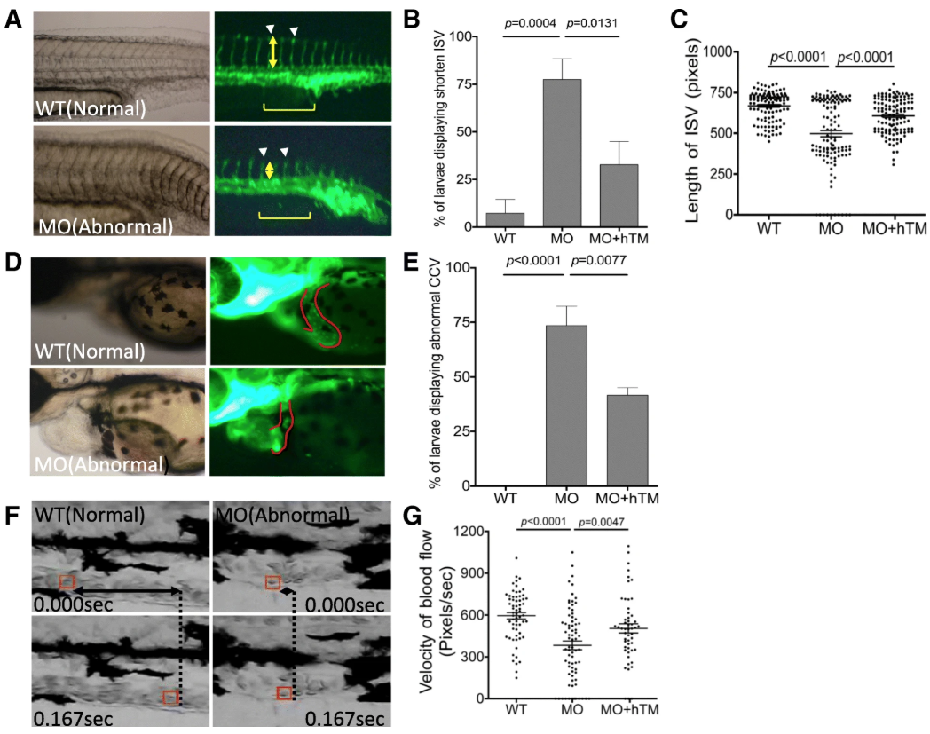
Fig. 4
The impact of zTM-b knockdown to vessels formation. Tg ( fli 1:eGFP) embryos were injected with TM-b MO with/without co-injecting plasmids encoding human TM, as described in Material and Methods, and observed for the development of ISV at 28 hpf ( a- c) and CCV at 56 hpf ( d, e). The percentage of morphants displaying anomaly in ISV (arrowheads) and CCV (CCV boundary circled by solid line) were also recorded. The extent of incomplete extension observed in ISV was quantified by calculating the length (yellow double-headed arrows) of five ISV singlets (brackets) immediately adjacent to cloaca for each morphant. f, g Larvae at 3 dpf were video-recorded laterally for the trunk area with anterior to the right for blood flow. A single red blood cell (boxed in red) inside the caudal vein was traced and calculated for its traveling distance (F; double-headed arrows) and velocity ( g) by analyzing the serial video still frames within 0.167 s. Presented are the results collected from at least three independent experiments including total 16–35 larvae for each experimental group. WT, wild-type embryos; MO, zTM-b morphants; hTM, human thrombomodulin