
|
Fig. 6
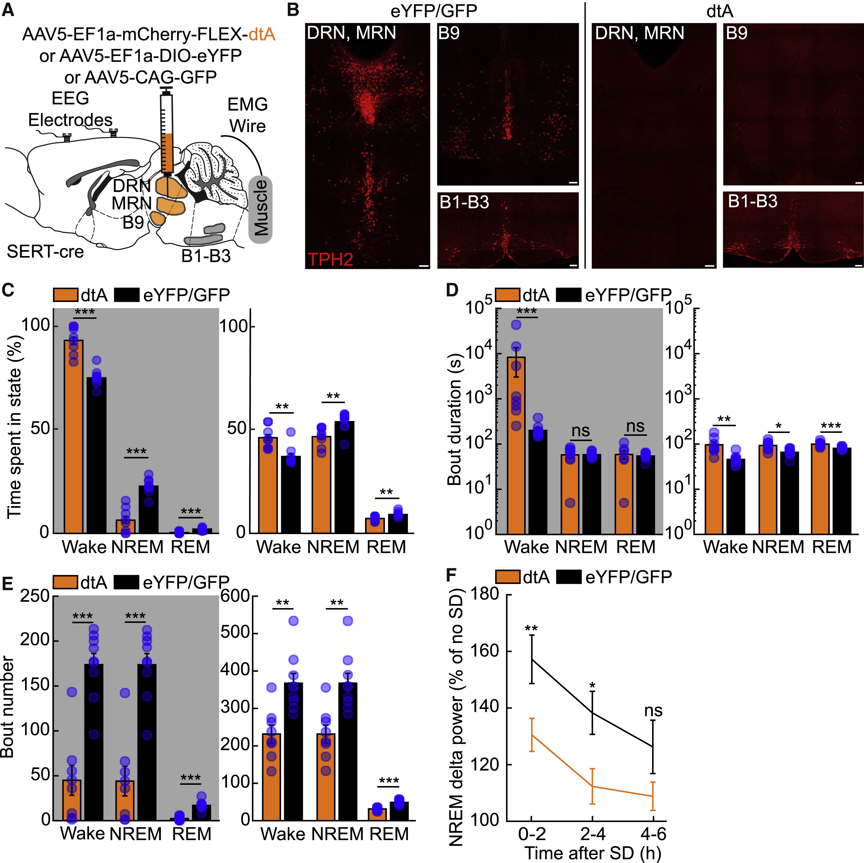
Ablation of Murine Raphe Leads to Increased Wakefulness and Impairs the Homeostatic Response to Sleep Deprivation
(A) Experimental setup.
(B) Representative images of TPH2+ neurons (red) from SERT-cre mice injected with either AAV5-EF1a-DIO-eYFP (left) or AAV5-EF1a-mCherry-FLEX-dtA (right). Scale bar, 100 μm.
(C) Percentage of time spent in the wake, NREM, and REM states during the dark (gray shading) and light phase from 24 h polysomnographic recordings.
(D and E) Duration (D) and number (E) of wake, NREM, and REM bouts during the dark and light phase.
(F) Change in delta power across NREM episodes in animals subjected to 6 h of sleep deprivation (SD) at the beginning of the light phase compared to undisturbed sleep from previous light phase (no SD).
n = 8 for B5-B9SERT-dtA, n = 9 for B5-B9SERT-eYFP or B5-B9GFP, two-sided Wilcoxon rank-sum test. ns p > 0.05, ∗p < 0.05, ∗∗p < 0.01, ∗∗∗p < 0.001.