
|
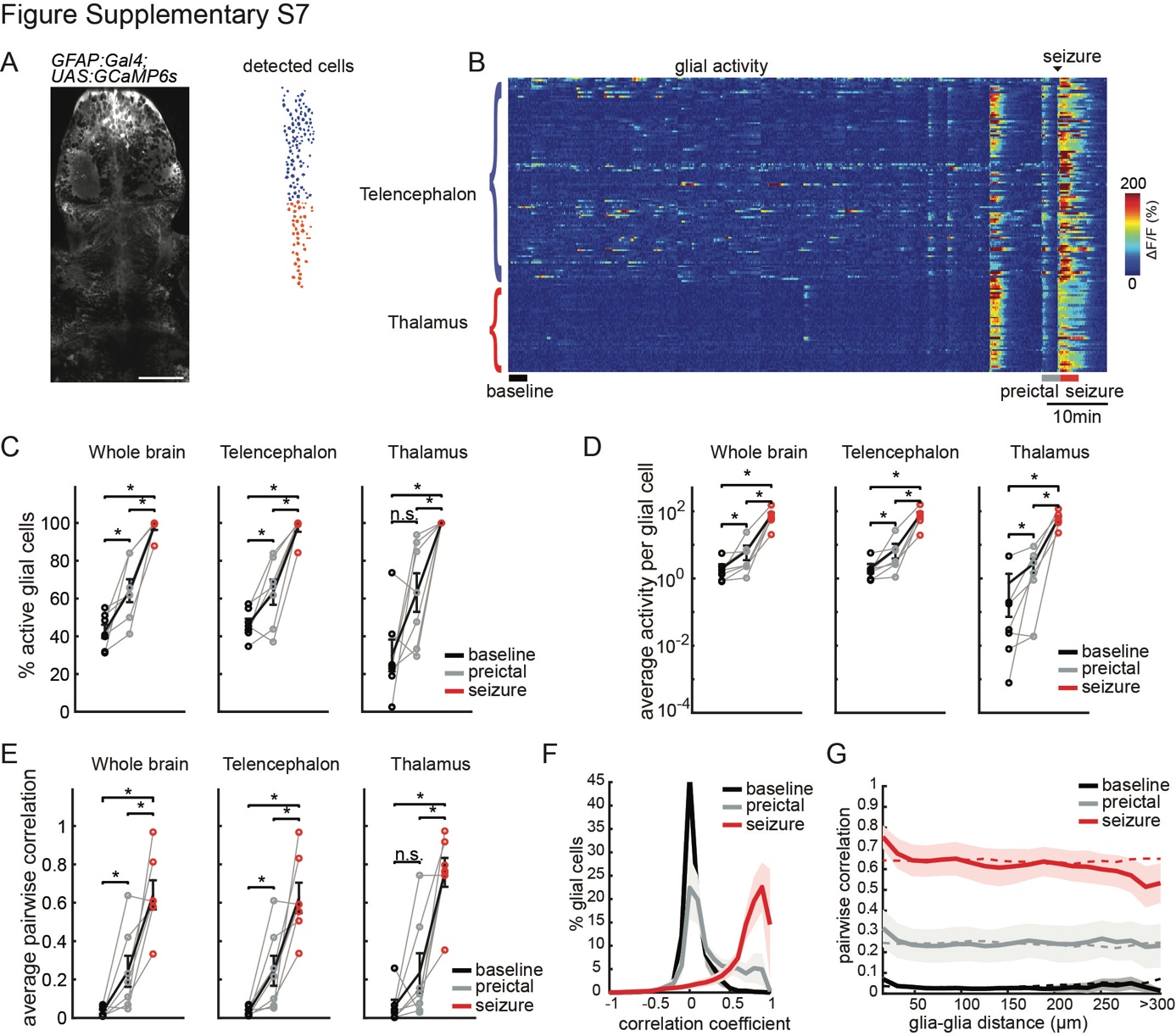
Fig. S7
Pilocarpine-induced seizures are preceded by strong glial activity and synchrony, similar to pentylenetetrazole (PTZ)-induced seizures
A) An optical section of a zebrafish larva expressing GCaMP6s in GFAP positive radial glia, obtained by two-photon microscopy, dorsal view (left). Individual glial cells (right) in color-coded brain regions: telencephalon (blue), and thalamus (red). White bar reflects 50 μm.
B) Activity of individual glial cells (ΔF/F) over time, organized by brain region. Application of 60 mM pilocarpine was done at the beginning of the recording. Baseline is indicated for a time period preceding strongly correlated activity. Warmer colors indicate stronger activity.
C) Percentage of active glial cells (>3stdbaseline) in the whole brain and per brain area during baseline (black), preictal (gray) and seizure (red) periods.
D) Average activity of the active glial cells, defined by the area under the curve of the ΔF/F trace, in the whole brain and per brain area.
E) Average pairwise Pearson’s correlation across the whole brain, and within individual brain regions.
F) Histogram representing the distribution of all correlation coefficients between glial cells from all animals.
G) Relation between pairwise Pearson’s correlation of glial activity and the distance between each glia pair. Dotted lines represent the results when glial locations are shuffled. n=7 fish. Shaded regions and error bars represent the s.e.m. (*p= <0.05, ns= not significant, Wilcoxon signed-rank test).