
|
Fig. S5
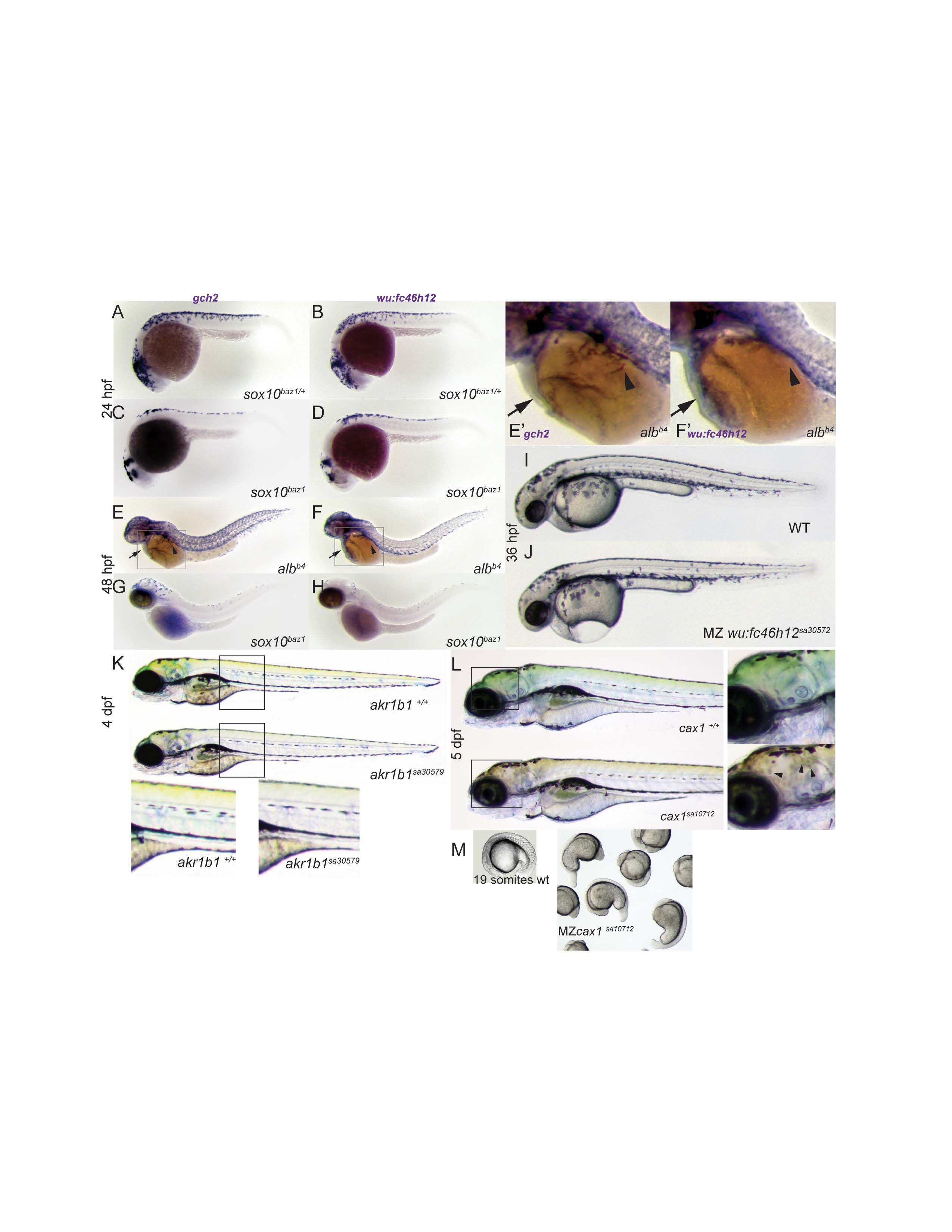
(A-H) Whole mount in situ analysis of wu:fc46h12 and gch2 as a pigment cell comparison. sox10baz/+ heterozygotes embryos as sibling controls (A-B) and mutant sox10baz1embryos at 24 hpf (C-D). At 48 hpf in situs were carried out on albino embryos to serve as wild-type controls (E-F) with arrows indicating the heart and arrow heads the dorsal aorta. A blow up of this region can be found in E’-F’. (G-H) Expression of gch2 and wu:fc46h12 at 48 hpf in sox10baz1 mutants. I-J Wild-type and MZwu:fc46h12sa30587 embryos at 36 hpf with oedema around the forming heart (J). (K) Wild-type sibling and mutant akr1b1sa30579at 4 dpf with mutant larvae presenting a reduction of yellow colour produced by xanthophores. Magnifications indicated with a black box. (L) Wild-type sibling and mutant cax1sa10712 larvae at 5 dpf. Close ups indicated by black boxes around the head show dull yellow colour and abnormal cell morphology in mutants (arrow head). (M) MZcax1sa10712phenotype at 19 somite stage.