
|
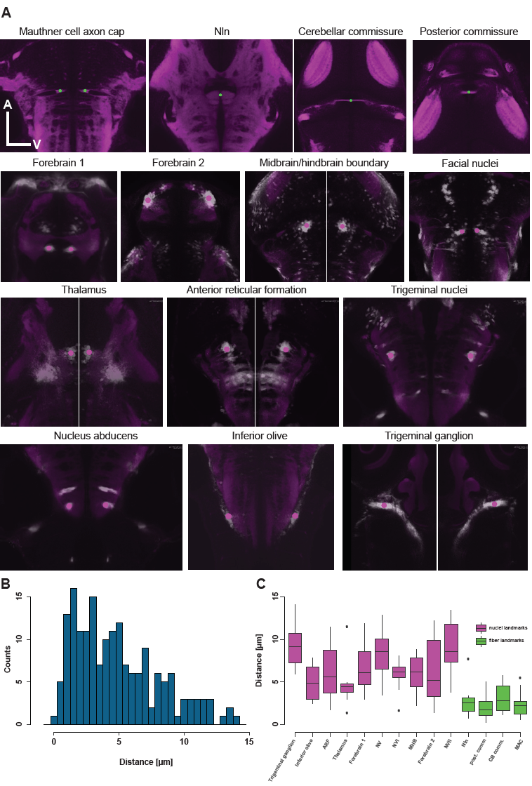
Fig. S3
Location of fiducial points used for measurement of registration accuracy.
(A) Single optical planes throughout the SYP-standard brain as well as through multiple ,different average Gal4-pattern indicating the location of the fiducial points (fiber landmarks are in green and nuclei landmarks are in magenta). (B) Histogram of all distances measured between fiducial points aligned to the standard brain and their respective counterpart in the standard brain. (C) Boxplot indicating the alignment accuracy for the individual fiducial points. Magenta refers to nuclei (Gal4-pattern) based landmarks and green to fiber (SYP-pattern) based landmarks.