
|
Fig. S5
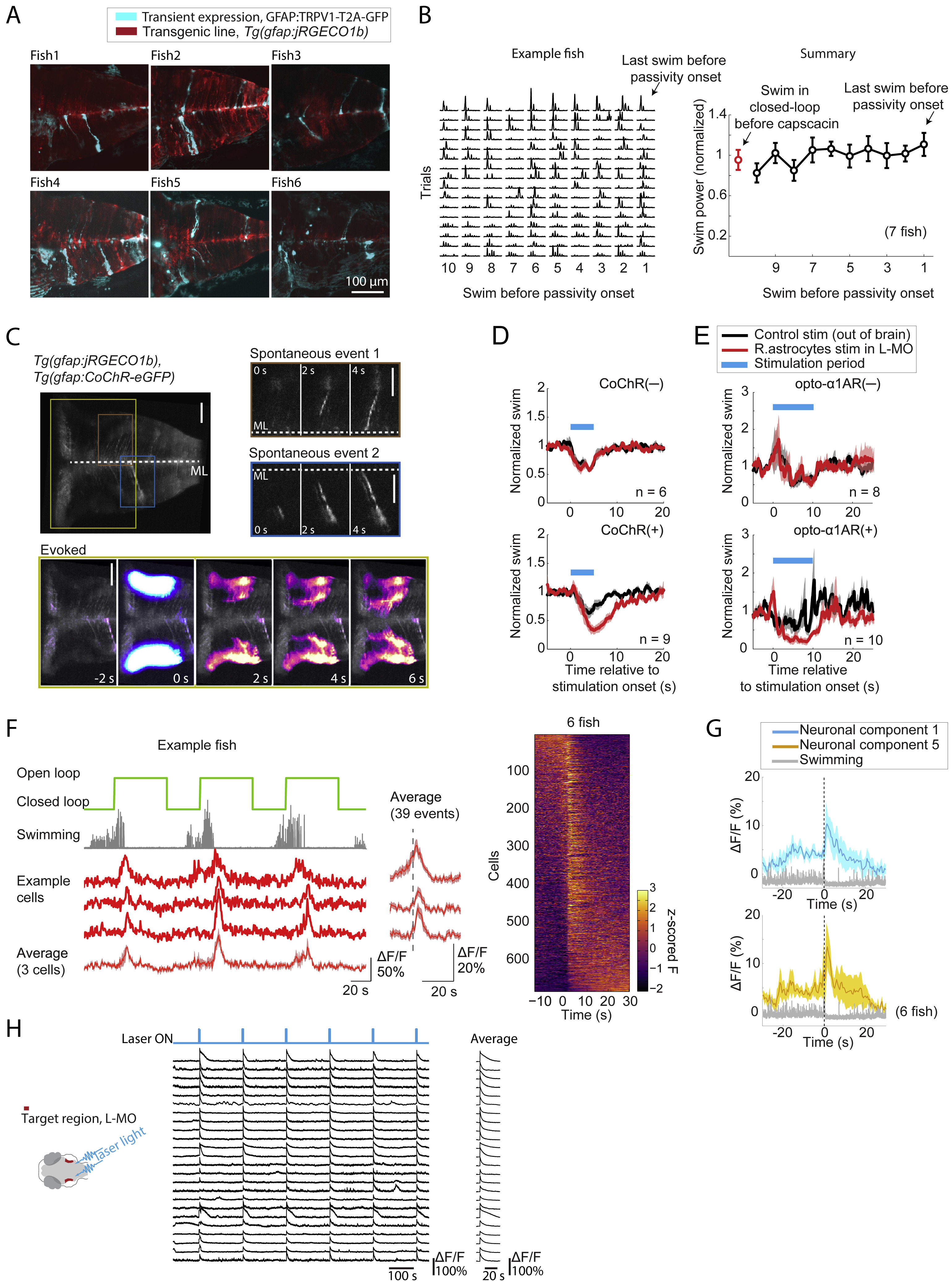
Details of Chemogenetic and Optogenetic Activation of Radial Astrocytes, Related to Figure 5
(A) Sparseness of expression of TRPV1. Maximum intensity projections along the dorsal-ventral axis of hindbrain volumes acquired with a light sheet microscope show that TRPV1 (Chen et al., 2016) channels (introduced into the fish by injection of a gfap:TRPV1-T2A-GFP, plasmid into Tg(gfap:jRGECO1b) transgenic embryos) were sparsely expressed in radial astrocytes. Expression was seen in about 10-50 astrocytic cells per animal, but opening of the TRPV1 channel with capsaicin activated a much wider population of glial cells (at least hundreds, likely through communication between radial astrocytes). Expressing cells showed the morphology of hindbrain radial astrocytes, with somata along the midline and processes extending laterally and then ramifying. (Note, the images show some contamination from skin autofluorescence. Also note, the jRGECO1b expression was more patchy than usual, likely because the co-injected Tol2 transposase removed the jRGECO1b transgene from the genome of some cells.)
(B) Artificially activating radial astrocytes induces passivity without increasing swim vigor, consistent with a role for radial astrocytes in specifically suppressing swimming. Passivity was caused in closed-loop by applying capsaicin to fish that expressed TRPV1 (Chen et al., 2016) in radial astrocytes. In the swim bouts just prior to TRPV1-mediated passivity, swim vigor did not increase as it did during futility-induced passivity (Figure S1C) – rather, swim vigor stayed at a similar level as swims in closed-loop without capsaicin, showing that radial astrocytes caused passivity independently of increases in swim vigor. In example fish (left panel), each row represents the final 10 swims of an active period before the fish switched to a passive state. Error bars, SEM.
(C) Optogenetic stimulation during functional imaging of radial astrocytes shows that exciting these cells can cause spatially propagating calcium activity. Top row: after optogenetic excitation of astrocytic processes in the L-MO, a wave of calcium can be seen propagating from the site of excitation to the astrocytic cell bodies located along the midline. The time course of evoked activity resembles spontaneous activity of single astrocytic cells (top right), which suggests that optogenetic stimulation is sufficient to drive an endogenous calcium-dependent excitatory pathway in radial astrocytes. Scale bars, 20 μm.
(D and E) Optogenetic stimulation of L-MO astrocytic processes suppresses swimming. (D) Top: CoChR-negative controls showed no difference in behavior when the L-MO was stimulated or light was targeted outside of the brain (targeted to the ear, chosen because it is very close to the L-MO but outside the brain). A small response to the blue light was present if either the L-MO or the ear was targeted. Bottom: The same experiment in animals expressing CoChR in radial astrocytes showed a difference in behavior, with L-MO stimulation leading to a prolonged suppression of swimming. In total 30 pulses with a width of 10 ms and inter-pulse-interval of 90 ms were delivered for each stimulation (Methods). Shading represents SEM (E) Stimulation in Opto-α1-AR expressing animals similarly showed a suppression of swimming. A continuous 10 s of light stimulation was delivered for each stimulation (Methods). Shading represents SEM.
(F) GABAergic neurons in L-MO activated during switches to passivity. Left, fish switching to the passive state after entering open-loop, with activity increases in GABAergic neurons in L-MO at the time of switches. Right, summary of all GABAergic neurons in L-MO from 6 fish. Cells are ordered by the center of mass along the time axis. Shading represents SEM.
(G) Capsaicin activation of astrocytic calcium causes an increase in neuronal activity in L-MO neurons, consistent with the activation of GABAergic neurons in L-MO after astrocytic stimulation (Figure 5R). Shading represent SEM.
(H) Optogenetically stimulating radial astrocytes causes increases in neuronal activity in L-MO neurons. Shading represents SEM.
Reprinted from Cell, 178(1), Mu, Y., Bennett, D.V., Rubinov, M., Narayan, S., Yang, C.T., Tanimoto, M., Mensh, B.D., Looger, L.L., Ahrens, M.B., Glia Accumulate Evidence that Actions Are Futile and Suppress Unsuccessful Behavior, 27-43.e19, Copyright (2019) with permission from Elsevier. Full text @ Cell