
|
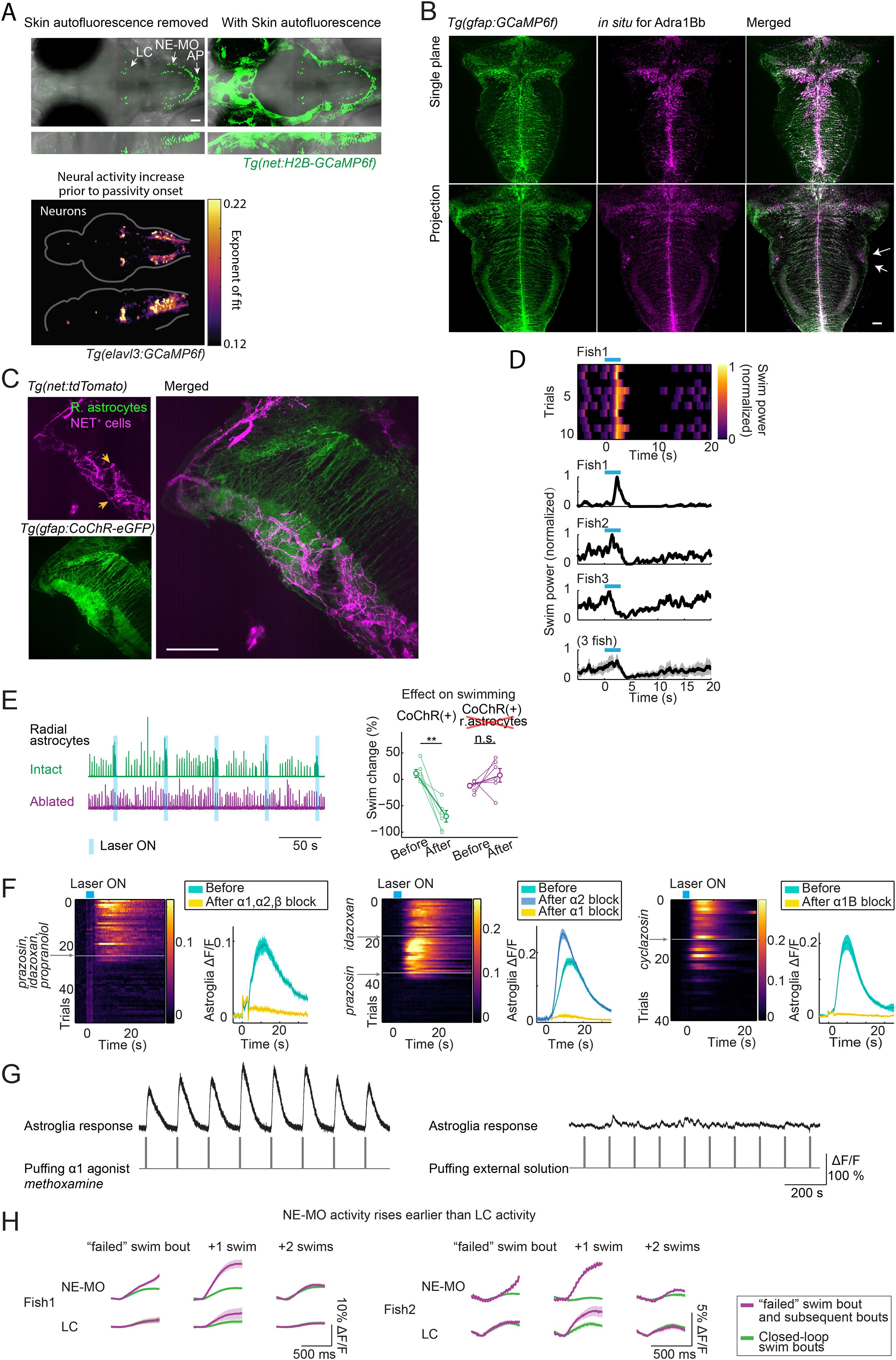
Fig. S6
Calcium Signals in Noradrenergic Neurons Increase before Switches to Passivity, Noradrenergic Receptors Are Highly Expressed and Co-localized with Radial Astrocytes in the Lateral Hindbrain, Activation of Noradrenergic Neurons Can Elicit a Transient Increase in Motor Vigor, and Coding for Swim Failures in Different NE Nuclei, Related to Figures 6 and 7
(A) Top: The nuclei of noradrenergic (NE) neurons (Farrar et al., 2018) are labeled in Tg(net:H2B-GCaMP6f) fish. Bottom: The neurons which increase their activity fastest prior to passivity onset overlap with the regions where the somata of noradrenergic cells, including the locus coeruleus (LC) and the NE cluster of the medulla oblongata (NE-MO), are located. Imaged line in the bottom panel is Tg(elavl3:GCaMP6f). For easier visualization of noradrenergic cells, skin autofluorescence was removed manually in the top-left panel.
(B) Adrenergic receptor 1Bb expression visualized through in situhybridization. The lateral hindbrain (L-MO) showed high levels of adra1Bb receptor expression (arrows). The receptors can also be seen to line the astrocytic processes that run medial-lateral, consistent with RNA-seq results (see Table S2). The projection is over a 40 μm thick section.
(C) Maximum intensity projection over 50 μm at the level of the L-MO of a Tg(gfap:CoChR-eGFP); Tg(net:tdTomato) fish that was expanded 4× through expansion microscopy to show a dense network of noradrenergic fibers expressing the norepinephrine transporter (NET) intertwined with glial processes in the L-MO. The section also shows the cell bodies of noradrenergic neurons (yellow arrows) located at the along the length of the medulla showing that indeed, noradrenergic neurons of the medulla oblongata (NE-MO) project to the L-MO.
(D) Photostimulating NE-MO can briefly potentiate swimming. In closed-loop, optogenetically stimulating NE-MO first increased swim vigor before it suppressed swimming. Top, single trials from an example fish. Bottom, mean swim power change from 3 example fish and the average from these 3 fish. Shaded region represents SEM.
(E) Photostimulating NE-MO no longer suppresses swimming after ablation of radial astrocytes in L-MO.
(F) Blocking α1 adrenergic receptors, but not α2 adrenergic receptors, abolishes the activation of radial astrocytes in L-MO by NE-MO stimulation. Optogenetic stimulation of NE-MO activates radial astrocytes in L-MO, and this activation was abolished by either adding a mixture of antagonists targeting multiple subtypes of adrenergic receptors (left, prazosin for α1, idazoxan for α2, propranolol for β), by an antagonist of α1 adrenergic receptors alone (middle), or by an antagonist of α1B adrenergic receptors alone (right), but not by an antagonist of α2 adrenergic receptors (middle). Shading represents SEM.
(G) Puffing methoxamine, an agonist of the α1 adrenergic receptor, triggered calcium responses in radial astrocytes in L-MO (left). Puffing external solution didn’t trigger a response (right).
(H) Calcium activity in the NE-MO represents swim failures (Figure 7A). Calcium activity in the LC also represents swim failures, but only on the subsequent swim bout; this response may be downstream of the response in the NE-MO. The signals were aligned by subtracting the fluorescence right before the swim onset. Shading represents SEM.
Reprinted from Cell, 178(1), Mu, Y., Bennett, D.V., Rubinov, M., Narayan, S., Yang, C.T., Tanimoto, M., Mensh, B.D., Looger, L.L., Ahrens, M.B., Glia Accumulate Evidence that Actions Are Futile and Suppress Unsuccessful Behavior, 27-43.e19, Copyright (2019) with permission from Elsevier. Full text @ Cell