
|
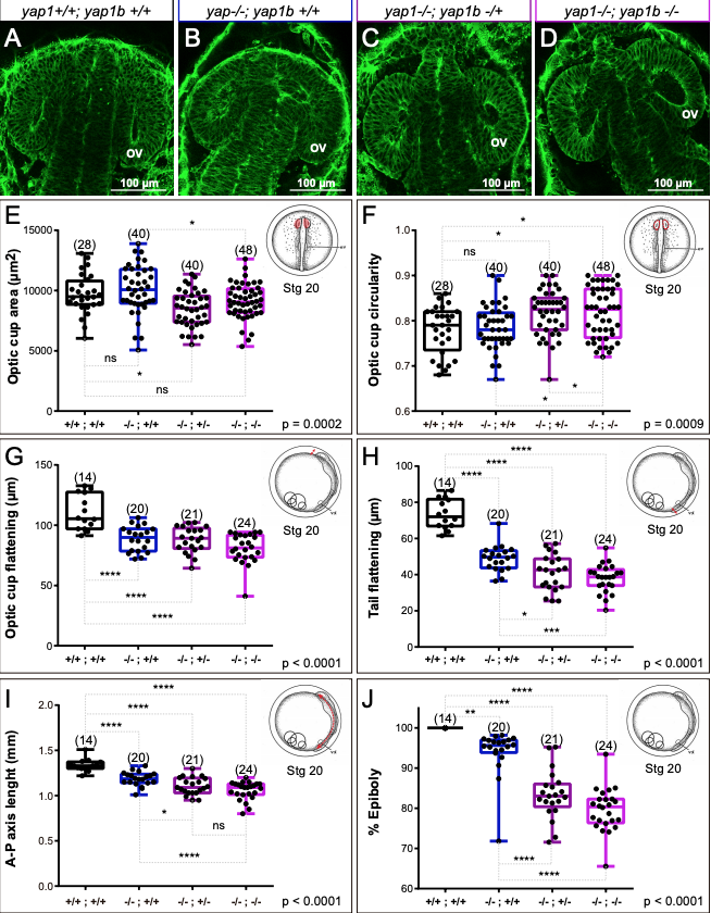
Fig. S13
Quantitative analysis of morphogenetic defects in yap1/yap1b single and double mutants. (A-D) Confocal sections of phalloidin stained embryos showing optic cup morphology in wild type, yap1 mutants, and yap1/yap1b double mutants. (E-J) Quantitative analysis of optic cup area (E), circularity (F), and flattening (G); as well as tail flattening (H), embryo axis lenght (I), and % of epiboly (J) in wild type and mutant embryos reveal enhanced tissue malformations in yap1/yap1b double mutants. One Way ANOVA analysis followed by Fisher's LSD test was used to evaluate statistical significance. ov = optic vesicle. Magnification bars are included in the figure.