
|
Fig. S5
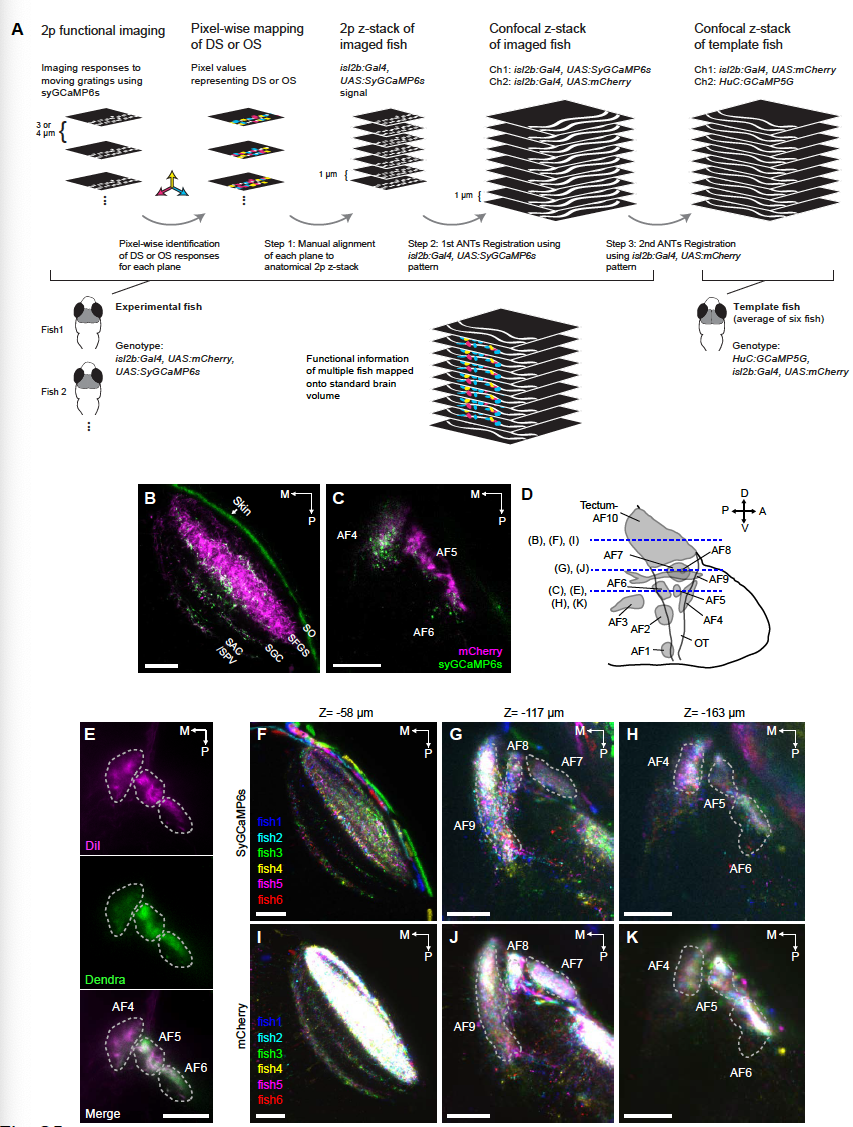
Image registration work flow to generate a 3D map of direction-selectivity, characterization of isl2b:Gal4, UAS:syGCaMP expression and overlay from image registration. (A) Schematic workflow for registering functional responses with anatomical structures. For clarity, only 3 preferred directions are represented here. See STAR Methods for details. (B, C) Subcellular localization of syGCaMP6s in the tectum/AF10 (B) and AF4, AF5 and AF6 (C) in isl2b:Gal4, UAS:syGCaMP6s, UAS:mCherry fish. Note that syGCaMP6s expression exhibits punctate signals in RGC terminals, in contrast to uniform mCherry signals in en passant RGC axon bundles. SO, stratum opticum; SFGS, stratum fibrosum et griseum superficiale; SGC, stratum griseum centrale; SAC, stratum album centrale. (D) Schematic illustration of AFs (modified from Burrill and Easter (1994)). Blue dotted lines indicate approximate z-planes shown in other panels of this figure. OT, optic tract. (E) Lipophilic dye DiI injection of the RGC axons in isl2b:Gal4, UAS:Dendra-kras fish. Note that the isl2b:Gal4 line labels most of RGCs projecting to AF4, AF5 and AF6. (F-K) Overlay of 6 different transgenic fish (isl2b:Gal4, UAS:syGCaMP6s, UAS:mCherry) that have been registered into a reference system (RGC standard brain based on isl2b:Gal4, UAS:mCherry). Z-position indicates the distance from the dorsal most surface of AF10. Note that both syGCaMP6s (F-H) and mCherry (I-K) patterns from 6 fish occupy conserved space in the registered volume. A, anterior; P, posterior; D, dorsal; V, ventral, M, medial. Scale bars represent 30 μm.