
|
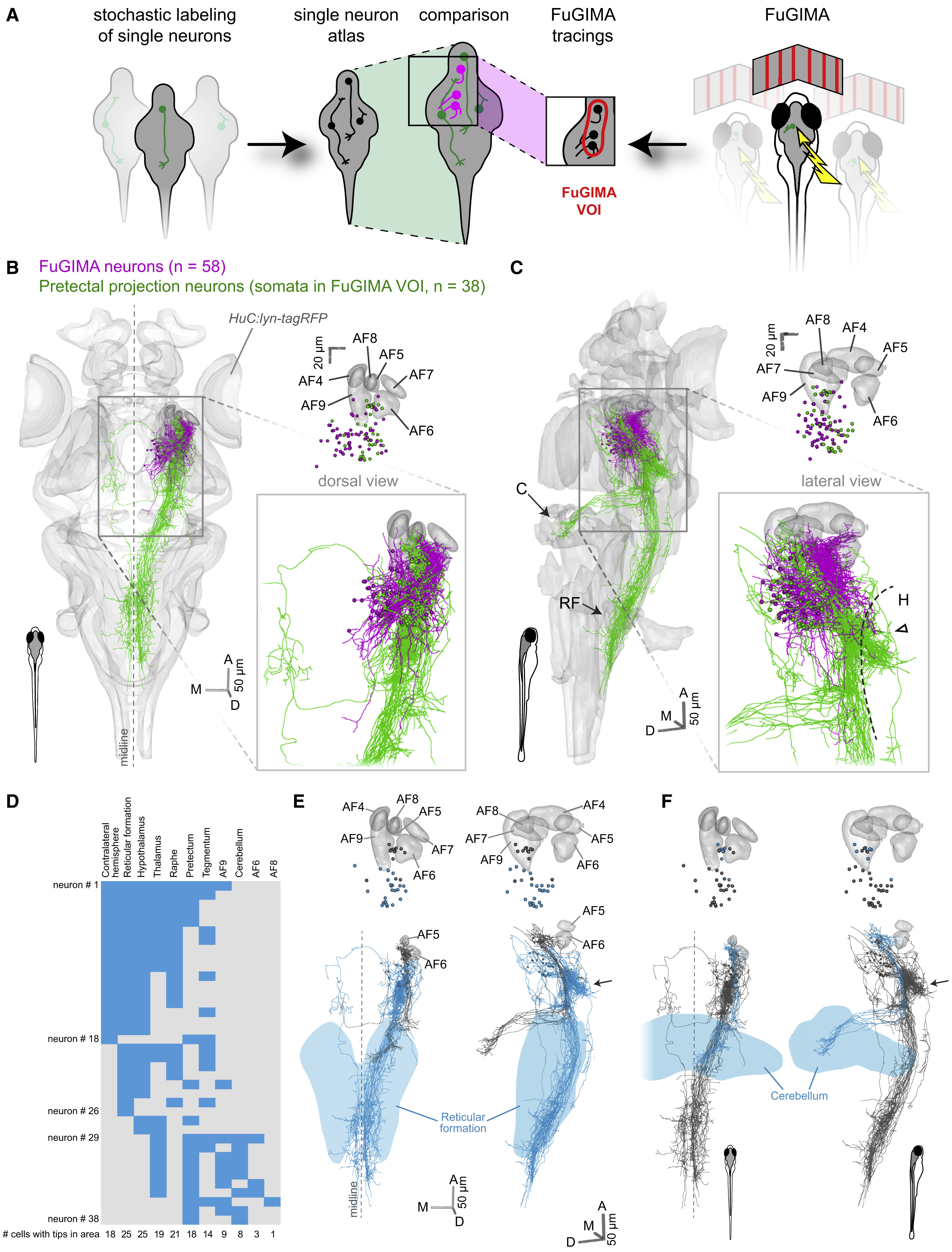
Fig. 6
Pretectal Projection Neurons Target the Cerebellum and Ventral Hindbrain
(A) Schematic illustrating the strategy to combine the single-neuron atlas of Kunst et al. (2019) and the FuGIMA dataset.
(B and C) 3D representation of the standard brain (HuC:lyn-tagRFP) together with all FuGIMA neurons (magenta, n = 58) as well as pretectal projection neurons (PPNs) (green, n = 38), chosen based on their soma location within the FuGIMA “volume-of-interest” (FuGIMA VOI) (Figure S7).
(B) (Left, dorsal view, top right) Dorsal view of cell bodies with AFs 4–9; (bottom right) detail of tracings.
(C) As (B) but lateral view (C, cerebellum; H, hypothalamus; RF, reticular formation; dashed line, dorsal border of hypothalamus; open arrowhead, dense branching of PPNs).
(D) Intersection analysis of PPNs with annotated brain areas, i.e., contralateral hemisphere, reticular formation, hypothalamus, thalamus, raphe, pretectum, tegmentum, AF9, cerebellum, AF6, and AF8. Each row represents one neuron; blue filled rectangles symbolize intersection with the annotated brain area.
(E) 3D rendering of intersection of PPNs with the reticular formation (blue, intersecting tracings [n = 25 of 38 PPNs]; gray, not intersecting PPNs; light blue, reticular formation; top, somata and AFs 4–9; bottom, tracings and AFs 5 and 6; left, dorsal view; right, lateral view; arrow, dense branching area in dorsal hypothalamus).
(F) As (E) but intersection of PPNs with the cerebellum (blue, intersecting tracings [n = 8 of 38 PPNs]; light blue, cerebellum).