
|
Fig. S1
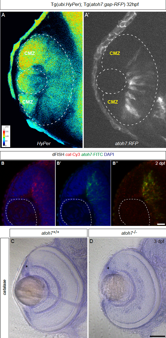
(A – B) H2O2 levels are decrease in differentiated cells, related to Figure 1. Tg(ubi:HyPer);Tg(atoh7:RFP) embryos imaged at 32 hours post-fertilization (hpf) show that as differentiation starts from the center of the tissue to its periphery, H2O2 levels are decrease where cells are differentiating and remain at high level at the periphery where the future ciliary marginal zone (CMZ) is forming. (B – B’’) At 2 dpf, catalase and atoh7 are expressed in the central retina transiently. (C – D) Catalase expression is independent from Atoh7 function in the retina. Comparative in situ hybridization at 3 dpf of catalase expression in wild type (C) and atoh7-/- mutant retinae (D) reveals that catalase is expressed in the absence of Atoh7 (asterisks, CMZ). Scale bar (B – B”) = 20 μm (C – D) = 100 μm.
Reprinted from Developmental Cell, 50(1), Albadri, S., Naso, F., Thauvin, M., Gauron, C., Parolin, C., Duroure, K., Vougny, J., Fiori, J., Boga, C., Vriz, S., Calonghi, N., Del Bene, F., Redox Signaling via Lipid Peroxidation Regulates Retinal Progenitor Cell Differentiation, 73-89.e6, Copyright (2019) with permission from Elsevier. Full text @ Dev. Cell