
|
Fig. S6
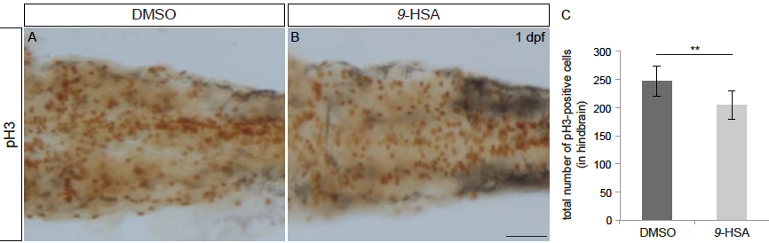
9-HSA exogenous administration leads to a decrease in proliferation in the hindbrain, related to Figure 5. pH3 staining (brown) labeling mitotic cells of whole-mount embryos injected with DMSO (control, A) and 9-HSA (B) at 1 day post-fertilization (dpf). (C) Quantification of the mitotic index in the hindbrain at 1 dpf reveals a significant decrease in the number of proliferative cells in the hindbrain of 9-HSA -treated compared to DMSO treated embryos (n = 7 for DMSO and n = 9 for 9-HSA –treated. Average number of pH3- positive cells in DMSO-treated hindbrain = 246, SD ± 26 and average number of pH3- positive cells in 9-HSA –treated hindbrain = 203, SD ± 25; p-value: 0.005). ** p-value < 0.01. Scale bar, 50 μm.
Reprinted from Developmental Cell, 50(1), Albadri, S., Naso, F., Thauvin, M., Gauron, C., Parolin, C., Duroure, K., Vougny, J., Fiori, J., Boga, C., Vriz, S., Calonghi, N., Del Bene, F., Redox Signaling via Lipid Peroxidation Regulates Retinal Progenitor Cell Differentiation, 73-89.e6, Copyright (2019) with permission from Elsevier. Full text @ Dev. Cell