
Fig. S5
|
Fig. S5
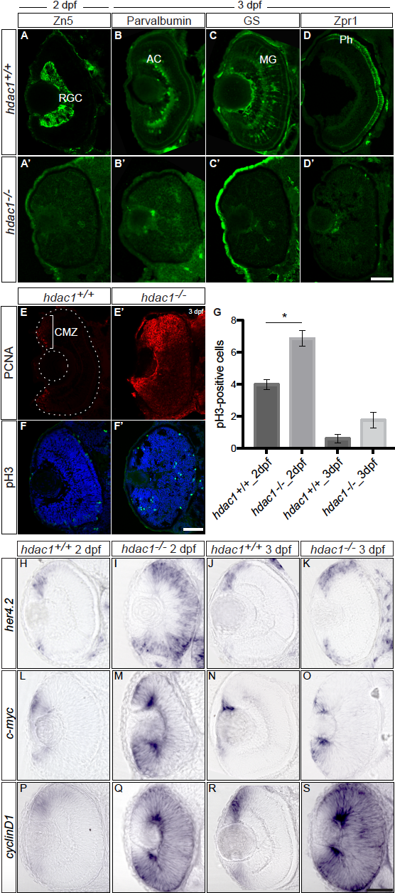
Hdac1 loss-of-function results in the activation of Notch and Wnt signaling pathways in the retina leading to an increase in the proliferation of retinal progenitor cells and differentiation defects for all retinal cell types, related to Figures 2, 3 and 4. (A – D’) Hdac1 loss-of-function results in a lack of differentiation in all cell types analyzed. (A – A’) Immunohistochemistry of anti-Zn5 (green) labeling retinal ganglion cells (RGC) on 2 dpf frontal retinal cryosections from wild type (hdac1+/+, A) and hdac1-/- mutant (A’) embryos. No RGC labeling could be observed in the central part of the retina at 2 dpf in 9-HSA -treated and hdac1-/- mutant retinae in comparison to wild type and DMSO-treated control retinae. (B – B’) Immunohistochemistry of anti-Parvalbumin (green) labeling amacrine cells and displaced amacrine cells (AC) on 3 dpf frontal retinal cryosections from wild type (hdac1+/+, B) and hdac1-/- mutant (B’) embryos. Similarly to RGCs, no amacrine cells or displaced amacrine cells could be observed in hdac1-/- mutant retinae in comparison to wild type retinae. (C – C’) Immunohistochemistry of anti-GS (green) labeling Müller glia cells (MG) on 3 dpf frontal retinal cryosections from wild type (hdac1+/+, C) and hdac1-/- mutant (C’) embryos shows no MG differentiation in hdac1-/- mutant retinae in comparison to the control retinae. (D – D’) Immunohistochemistry of anti-Zpr1 (green) labeling photoreceptor cells (Ph) on 3 dpf frontal retinal cryosections of wild type (hdac1+/+, D) and hdac1-/- mutant (D’) embryos. No Zpr1 labeling could be observed in hdac1-/- mutant retinae, while in wild type retinae Zpr1-positive Ph could be detected within the outer nuclear layer of the 3 dpf retinal tissue. Scale bars (A – D’) = 50 μm. (E – E’) Immunohistochemistry of anti-PCNA (red) labeling cells in S-phase of the cell cycle on 3 days post-fertilization (dpf) frontal retinal cryosections. Hdac1-/- retinae show an expanded PCNA labeling in the ciliary marginal zone (CMZ, bracket) and in the central retina in comparison to wild type sibling (hdac1+/+) retinae at 3 dpf. (F – F’) Immunohistochemistry of anti-pH3 (green) labeling cells in M-phase of the cell cycle on 3 dpf frontal retinal cryosections counterstained with the nuclear marker DAPI (blue). (G) The quantification of the number of pH3-positive cells in hdac1-/- in comparison to hdac1+/+ retinae shows a significantly higher number of mitotic cells in comparison to wild type retinae at 2 dpf (n = 3 retinae per condition, Unpaired Welch’s t-test, p-value = 0.0335 depicted as *). (H – S) Hdac1 loss-of-function results in the activation of Notch and Wnt signaling pathways in the retina. Expression analysis of her4.2, c-myc, and cyclinD1 at 2 days post-fertilization (dpf) in wild type siblings (hdac1+/+, respectively H, L, P), hdac1-/- mutant embryos (I, M, Q) and at 3 dpf (hdac1+/+, respectively J, N, R and hdac1-/-, respectively K, O, S). All the genes had increased expression in the retinae of hdac1-/- mutant in comparison to hdac1+/+ wild type siblings. Transcripts at 2 dpf could still be detected in the central part of hdac1-/- retinae. At 3 dpf, expression cyclinD1 expression was retained in the entire retinal tissue of hdac1-/- embryos in comparison to hdac1+/+ wild type siblings where cyclinD1 transcripts were restricted to the ciliary marginal zone (R – S). At this same stage, her4.2 and c-myc expressions were increased and expanded at the periphery of the tissue in the retinae of hdac1-/- embryos in comparison to hdac1+/+ wild type sibling retinae (K – J and O – N respectively). Scale bar = 50 μm.
Reprinted from Developmental Cell, 50(1), Albadri, S., Naso, F., Thauvin, M., Gauron, C., Parolin, C., Duroure, K., Vougny, J., Fiori, J., Boga, C., Vriz, S., Calonghi, N., Del Bene, F., Redox Signaling via Lipid Peroxidation Regulates Retinal Progenitor Cell Differentiation, 73-89.e6, Copyright (2019) with permission from Elsevier. Full text @ Dev. Cell