
|
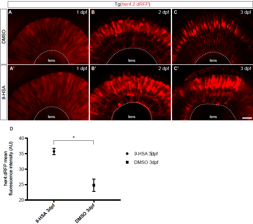
Fig. S4
Tg(her4.2:dRFP) embryos injected with 9-HSA display increased RFP expression from 2 to 3 days post-fertilization, related to Figure 3. Tg(her4.2:dRFP) embryos were injected at 1-cell stage with either 9-HSA or DMSO. The effect of the injection on the expression level of her4.2 was assessed at 1, 2 and 3 days post-fertilization (dpf). At 1 dpf, no difference could be observed between the retinae of 9-HSA and DMSO injected embryos (A – A’). At 2 and 3 dpf, an increased expression of her4.2:dRFP could be observed in the retinae of 9- HSA in comparison to DMSO injected embryos of the same developmental stage (B – C’). (D) Quantification of the her4:dRFP fluorescent intensity at 3 dpf showed a significant increase in 9-HSA injected retinae of the dRFP signal compared to the control DMSO injected retinae (35.71 SEM ± 1.038 AU for 9-HSA and 24.79 SEM ± 2.005 AU for DMSO injected embryos, p-value: 0.0168, unpaired Welch’s t-test). Scale bar (A – C’) = 20 μm.
Reprinted from Developmental Cell, 50(1), Albadri, S., Naso, F., Thauvin, M., Gauron, C., Parolin, C., Duroure, K., Vougny, J., Fiori, J., Boga, C., Vriz, S., Calonghi, N., Del Bene, F., Redox Signaling via Lipid Peroxidation Regulates Retinal Progenitor Cell Differentiation, 73-89.e6, Copyright (2019) with permission from Elsevier. Full text @ Dev. Cell