
|
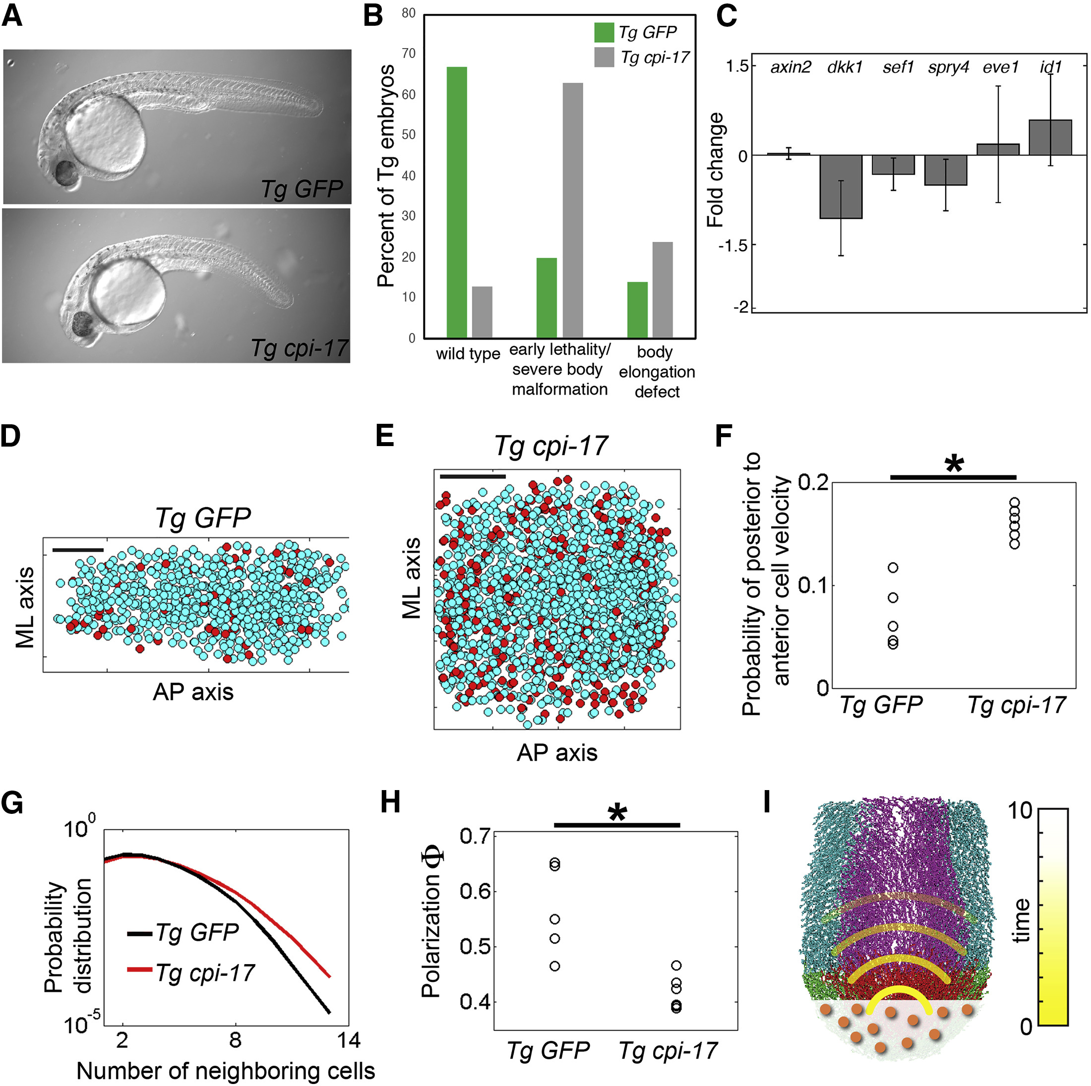
Fig. 6
Mechanical Information Propagation after Increasing Cell Contractility in the Tail Organizer
(A) Transient tbx6l transgenics expressing either GFP (control) or cpi-17 and GFP in the tail organizer.
(B) Tg tbx6l:GFP transgenics produce mostly normal embryos (3 experiments; n = 363) whereas Tg tbx6l:cpi-17 embryos (4 experiments; n = 416) exhibit a high frequency of early developmental defects and embryos with abnormal body elongation.
(C) Nascent transcription of Wnt, Fgf, and Bmp target genes was examined by qRT-PCR on pooled dissected tail buds. Expression in Tg tbx6l:cpi-17 tail buds was normalized to expression in Tg tbx6l:GFP controls. Data are averages from three independent experiments and display standard error.
(D and E) Snapshots of cells of the PNT in (D) a Tg tbx6l:GFP control (see Video S4) and (E) a Tg tbx6l:cpi-17 embryo (see Video S5). Red dots indicate cells with a posterior-to-anterior velocity. Scale bars in (D) and (E) represent 50 μm.
(F) The probability of posterior-to-anterior cell velocities in the PNT in five Tg tbx6l:GFP controls and six Tg tbx6l:cpi-17 embryos.
(G) Cell density in the PNT is displayed via the probability distribution of the number of neighboring cells. Each probability distribution is obtained by pooling the data from each experimental condition. These two probability distributions differ (p < 0.05, t test), and show that cell density is higher in Tg tbx6l:cpi-17 embryos.
(H) Global order of PNT cell velocities as measured by the polarization Φ.
(I) Mechanical information (yellow) extends the sphere of influence of the tail organizer beyond that of a secreted signaling molecule (orange).
Asterisks denote p < 0.05 (t test).
Reprinted from Developmental Cell, 49(6), Das, D., Jülich, D., Schwendinger-Schreck, J., Guillon, E., Lawton, A.K., Dray, N., Emonet, T., O'Hern, C.S., Shattuck, M.D., Holley, S.A., Organization of Embryonic Morphogenesis via Mechanical Information, 829-839.e5, Copyright (2019) with permission from Elsevier. Full text @ Dev. Cell