
|
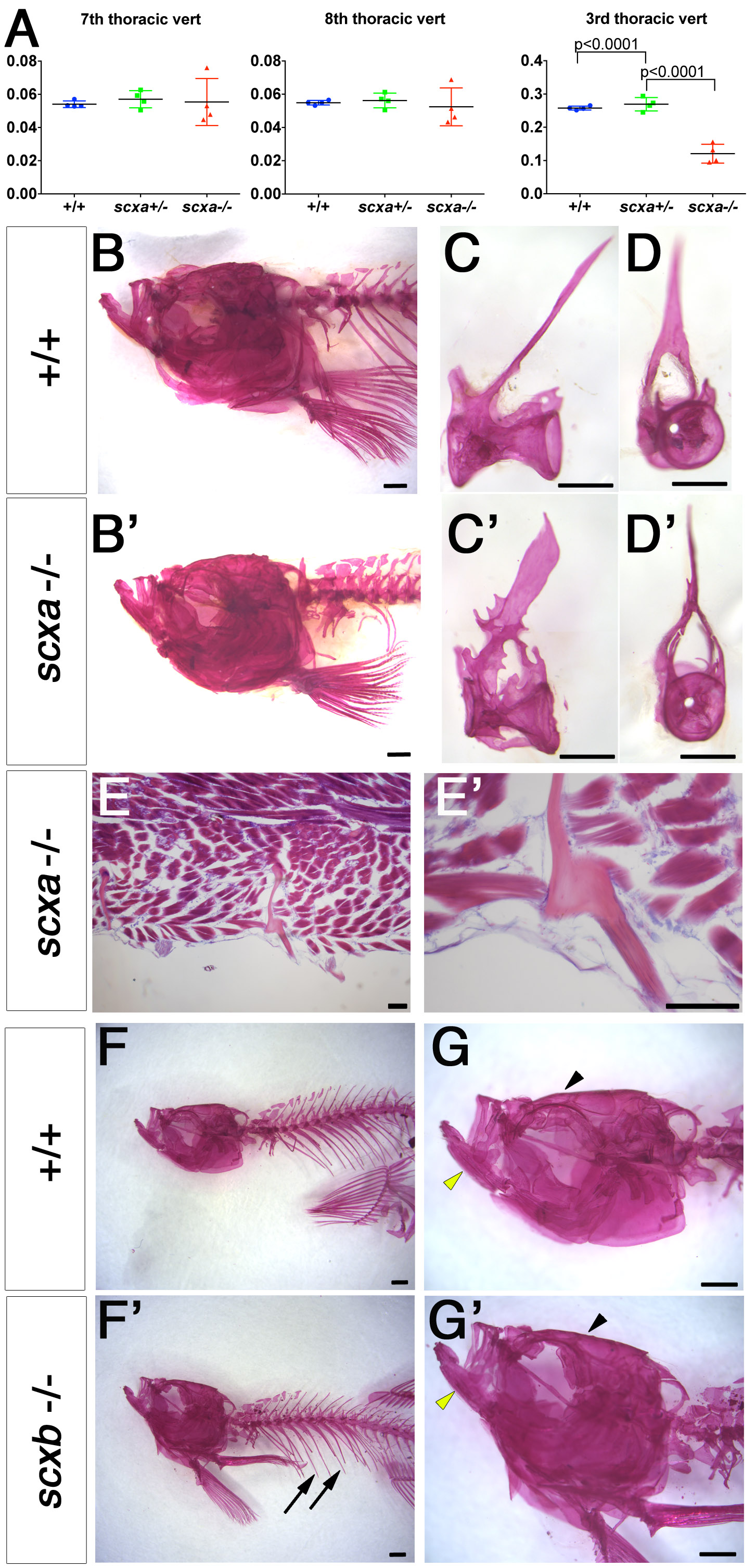
Fig. S5
Adult scxa mutants show skeletal defects in trunk but not in skull. A. Vertebral centrum volumes calculated from μCT scans of genotyped adults from a scxa+/- incross (n=3 per genotype) using the minimum volume possible around the neural canal, excluding (for 7th and 8th thoracic vertebrae) or including (for the 3rd thoracic vertebra) all processes, trabeculae, spines and ribs in a transverse view. Vertebrae centrum alone is similar between all genotypes, but when the above skeletal elements were added, volume in scxa-/- fish is significantly smaller than siblings. One-way ANOVA statistics with Tukey’s post-hoc test performed, p-values indicated. B-D’, F-G’. Alizarin Red staining for adult scxa-/- (B’-D’) and siblings (B-D) or scxb-/- mutants (F’,G’) and their siblings (F,G). Skull of scxa mutants appeared normal whereas staining in ribs was missing. Skull (arrowhead), jaw (yellow arrowhead) and ribs (arrows) looked normal in scxb mutants. Dissected thoracic 12th vertebrae from scxa-/- mutant and sibling are shown in lateral and frontal views for details of bony growth in arches (C’,D’ compared with C,D). E, E’. Adult scxa mutant zebrafish sagittal paraffin section stained with Hematoxylin and Eosin and Alcian blue showing fractured and healed rib fragment (magnified in E’). Scale bars, 100μm in E,E’, 1mm in B,B’,F,F’, 0.5mm in C-D’.